

Lipoid proteinosis
- PMID: 21887007
- PMCID: PMC3162866
- DOI: 10.4103/0973-029X.57675
Lipoid proteinosis
Abstract
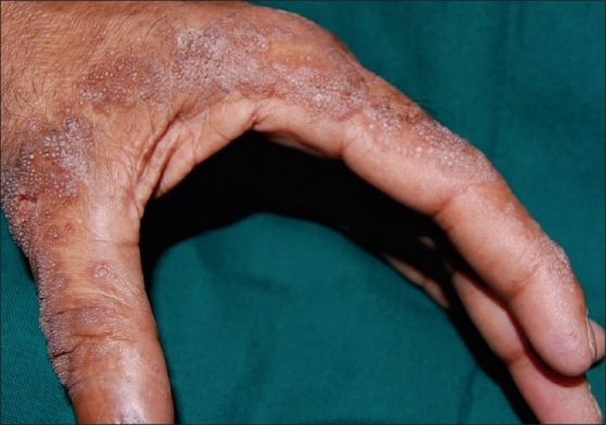
Lipoid proteinosis is a rare disorder with autosomal recessive inheritance, characterized by progressive deposition of hyaline material in the skin, mucous membrane, and different organs of the body, resulting in a multitude of clinical manifestations. A 62-year-old male presented with hoarseness of voice since infancy, eyelid beading, and generalized acneiform scars on the facial skin and extremities, and yellowish papules on his tongue and buccal mucose. The patient was diagnosed clinically as a case of Lipoid proteinosis, which was confirmed by skin and mucosal biopsy. The objective of the present work is to describe this rare entity, with approximately 250 cases found in medical literature. This case report also illustrates that Lipoid proteinosis may show protean clinical features and yet may remain undiagnosed for many years. This report will hopefully spawn further studies that will lead to early diagnosis.
Keywords: Acneiform scars; eyelid beading; hoarseness of voice; hyaline material; yellowish papules.
Conflict of interest statement
Figures


References
-
- Black MM. Lipoid Proteinosis, Metabolic and nutritional disorders. In: Champion RH, Burton JL, Burns DA, Breathnach SM, editors. Rook/ Wilkison/ Ebling Textbook of Dermatology. 6th ed. Oxford: Blackwell Science; 1998. pp. 2460–2.
-
- Touart DM, Sau P. Cutaneous deposition diseases. Part I. J Am Acad Dermatol. 1998;39:149–71. - PubMed
-
- Van Hougenhouck-Tulleken W, Chan I, Hamada T, Thornton H, Jenkins T, McLean WH, et al. Clinical and molecular characterization of Lipoid Proteinosis in Namaqualand, South Africa. Br J Dermatol. 2004;151:413–23. - PubMed
-
- Hamada T. Lipoid Proteinosis. Clin Exp Dermatol. 2002;27:624–9. - PubMed
-
- Hamada T, Mclean WHI, Ramsay M, Ashton GHS, Nanda A, Jenkins T, et al. Lipoid Proteinosis maps to 1q21 and is caused by mutations in the extracellular matrix protein 1 gene (ECM1) Hum Mol Genet. 2002;11:833–40. - PubMed
