

Meningiomas in children: A study of 18 cases
- PMID: 21887184
- PMCID: PMC3162790
- DOI: 10.4103/1817-1745.57322
Meningiomas in children: A study of 18 cases
Abstract
Background: Intracranial meningiomas are rare tumors in children accounting for 0.4-4.6% of all primary brain tumors in the age group of 0-18 years.
Objective: To retrospectively analyze the epidemiological profile, clinical features, radiological findings, type of excision, histopathological findings, and overall management profile of these patients.
Materials and methods: Eighteen consecutive cases of meningioma in patients under 18 years of age admitted and operated at our institute between the years 1974-2005 were included in this study.
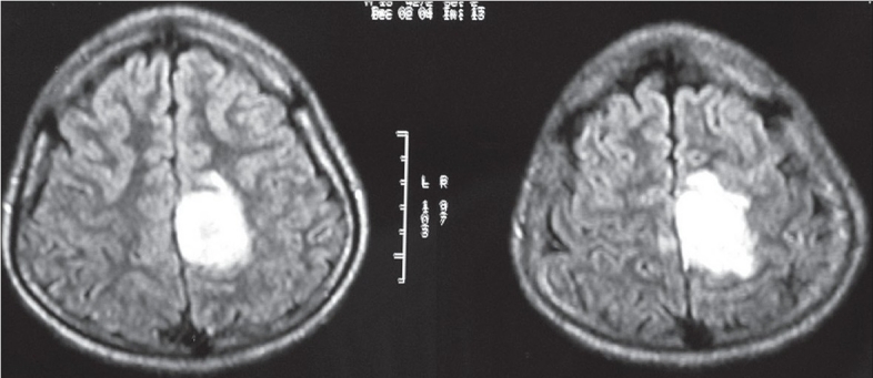
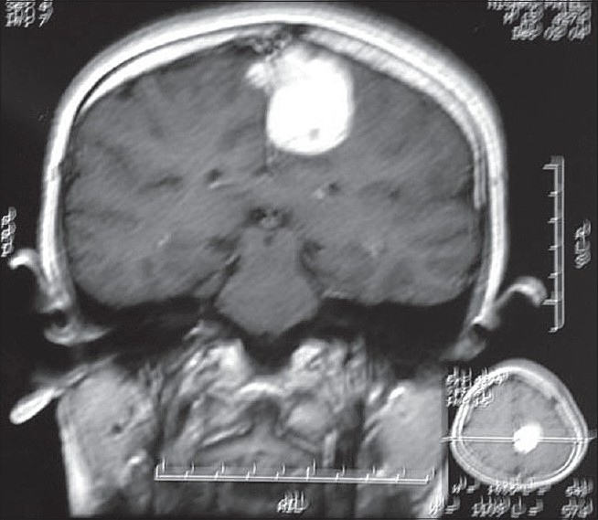
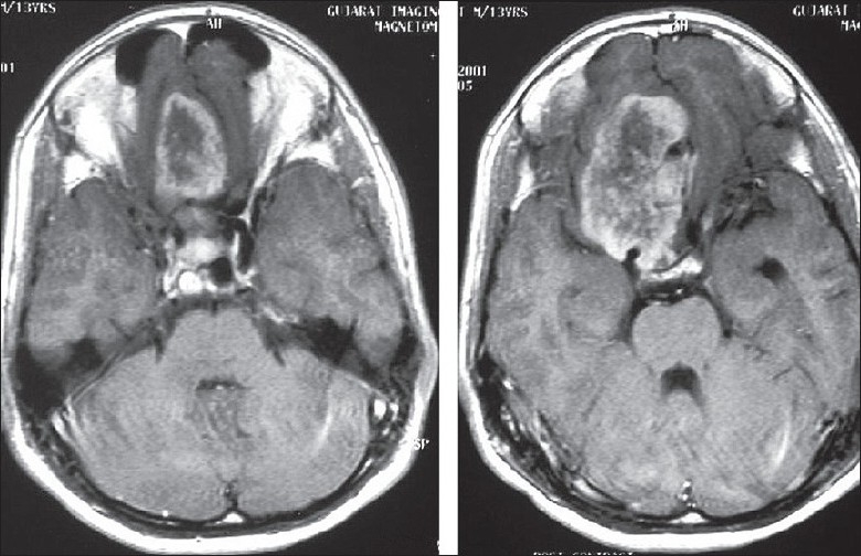
Results: The mean age of patient at presentation to our hospital was 12.81 years. The male to female ratio was 1.57:1. The median preoperative duration of symptoms was 1.2 years. An increased incidence was seen in patients with neurofibromatosis. Intraventricular and skull base locations were common. Total tumor excision was achieved in all cases.
Conclusion: A higher incidence of atypical and aggressive meningiomas is seen in children. Children with complete resection and a typical benign histology have a good prognosis.
Keywords: Meningioma; childhood meningioma; neurofibromatosis; pediatric neoplasms.
Conflict of interest statement
Figures
References
-
- Germano IM, Edwards MS, Davis RL, Schiffer D. Intracranial meningiomas of the first two decades of life. J Neurosurg. 1994;80:447–53. - PubMed
-
- Bodian M, Lawson D. Intracranial neoplastic diseases of childhood: A description of their natural history based on a clinico-pathological study of 129 cases. Br J Surg. 1953;40:368–92. - PubMed
-
- Cuneo HM, Rand CW. Vol. 15. Springfield IL, Charles C, Thomas: 1952. Brain tumors of childhood.
-
- French L. Intracranial and cranial tumor. In: Jackson P, Thompson F, editors. Pediatric Neurosurgery. Oxford UK: Blackwell; 1959. pp. 226–94.
-
- Keith HM, Craig WK, Kernohan JW. Brain tumors in children. Pediatrics. 1949;3:838–44. - PubMed
LinkOut - more resources
Full Text Sources
Research Materials
