

Pediatric craniofacial surgery for craniosynostosis: Our experience and current concepts: Part -1
- PMID: 21887189
- PMCID: PMC3162795
- DOI: 10.4103/1817-1745.57327
Pediatric craniofacial surgery for craniosynostosis: Our experience and current concepts: Part -1
Abstract
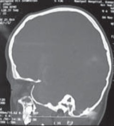
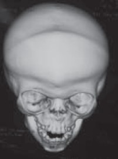
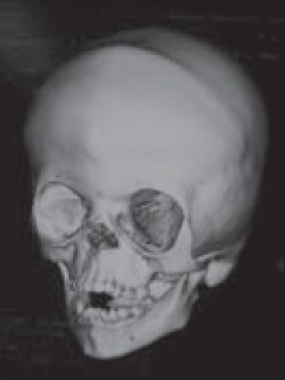
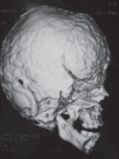
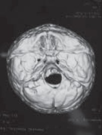
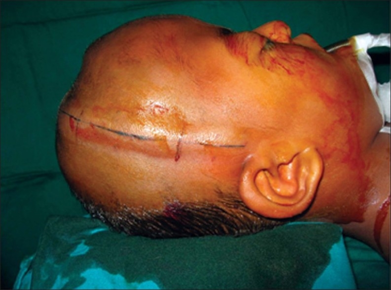
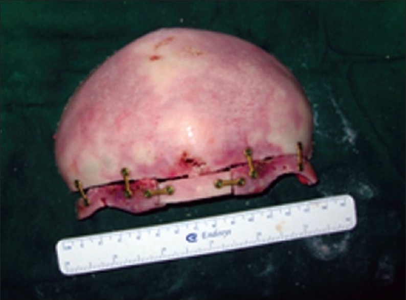
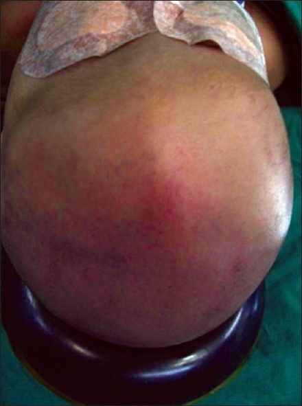
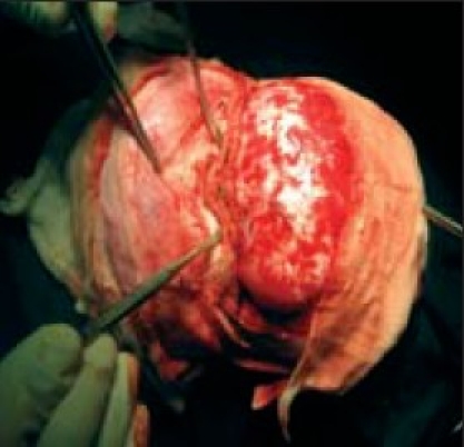
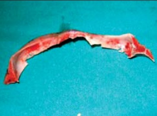
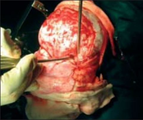
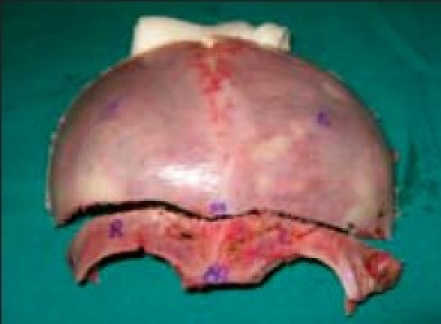
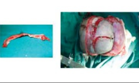
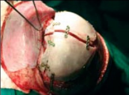
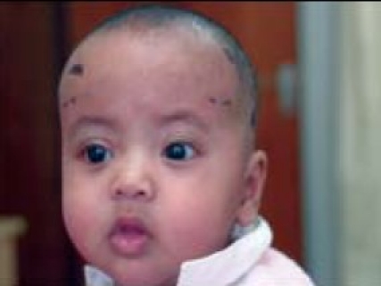
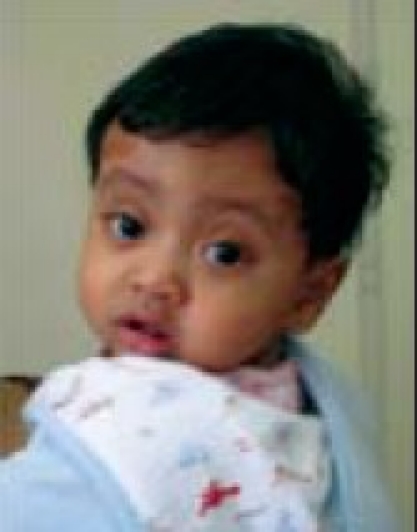
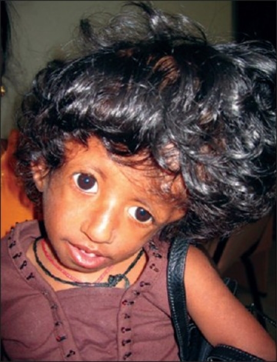
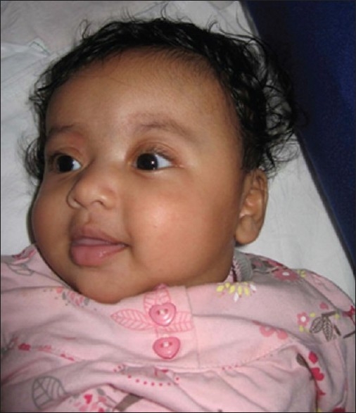
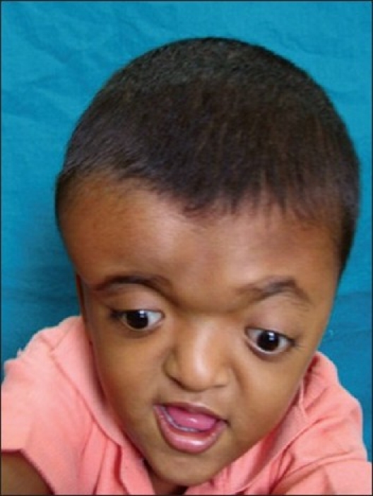
Craniostenosis is a disease characterized by untimely fusion of cranial sutures resulting in a variety of craniofacial deformities and neurological sequelae due to alteration in cranial volume and restriction of brain growth. This involves vault sutures predominantly, but cranial base is not immune. Association with a variety of syndromes makes the management decision complex. These children need careful evaluation by multiple specialists to have strategic treatment options. Parental counseling is an important and integral part of the treatment. Recent advancements in the surgical techniques and concept of team approach have significantly enhanced the safety and outcome of these children. We had an opportunity of treating 57 children with craniostenosis in the last 15 years at our craniofacial service. Out of them, 40 were nonsyndromic and 17 were syndromic variety. We describe our successful results along with individualized operative technical modifications adopted based on the current understanding of the disease.
Keywords: Nonsyndromic craniostenosis; operative results; pediatric craniofacial surgery.
Conflict of interest statement
Figures

Similar articles
-
Pediatric craniofacial surgery for craniosynostosis: Our experience and current concepts: Parts -2.J Pediatr Neurosci. 2009 Jul;4(2):100-7. doi: 10.4103/1817-1745.57328. J Pediatr Neurosci. 2009. PMID: 21887190 Free PMC article.
-
Evaluation and management of nonsyndromic craniosynostosis.Acta Paediatr. 2011 Sep;100(9):1185-94. doi: 10.1111/j.1651-2227.2011.02299.x. Epub 2011 May 5. Acta Paediatr. 2011. PMID: 21457300 Review.
-
Incidence of Cranial Base Suture Fusion in Infants with Craniosynostosis.Plast Reconstr Surg. 2018 Apr;141(4):559e-570e. doi: 10.1097/PRS.0000000000004238. Plast Reconstr Surg. 2018. PMID: 29595734
-
Multidisciplinary care of craniosynostosis.J Multidiscip Healthc. 2017 Jul 6;10:263-270. doi: 10.2147/JMDH.S100248. eCollection 2017. J Multidiscip Healthc. 2017. PMID: 28740400 Free PMC article. Review.
-
Skull vault growth in craniosynostosis.Childs Nerv Syst. 2005 Oct;21(10):861-70. doi: 10.1007/s00381-004-1112-2. Epub 2005 Mar 25. Childs Nerv Syst. 2005. PMID: 15791470 Review.
Cited by
-
Split cranial bone grafting in children younger than 3 years old: debunking a surgical myth.Plast Reconstr Surg. 2014 Jun;133(6):822e-827e. doi: 10.1097/PRS.0000000000000222. Plast Reconstr Surg. 2014. PMID: 24867741 Free PMC article.
-
Locally affine diffeomorphic surface registration for planning of metopic craniosynostosis surgery.Med Image Comput Comput Assist Interv. 2017 Sep;10434:479-487. doi: 10.1007/978-3-319-66185-8_54. Epub 2017 Sep 4. Med Image Comput Comput Assist Interv. 2017. PMID: 29527598 Free PMC article.
-
Radiological Diagnosis of Crouzon Syndrome: A Case Study.Cureus. 2024 Jun 17;16(6):e62564. doi: 10.7759/cureus.62564. eCollection 2024 Jun. Cureus. 2024. PMID: 39027794 Free PMC article.
-
Insights into craniosynostosis management in low- and middle-income countries: A narrative review of outcomes, shortcomings and paediatric neurosurgery capacity.SAGE Open Med. 2024 Jan 18;12:20503121241226891. doi: 10.1177/20503121241226891. eCollection 2024. SAGE Open Med. 2024. PMID: 38249946 Free PMC article. Review.
-
The use of a single-piece bone flap for cranial reshaping in anterior craniosynostosis patients: clinical experience and a description of a novel technique.Maxillofac Plast Reconstr Surg. 2022 Jan 5;44(1):2. doi: 10.1186/s40902-021-00332-4. Maxillofac Plast Reconstr Surg. 2022. PMID: 34985605 Free PMC article.
References
-
- Persing JA, Babler WJ, Jane JA, Duckworth PF. Experimental unilateral coronal synostosis in rabbits. Plast Reconstr Surg. 1986;77:369–76. - PubMed
-
- Mooney MP, Losken HW, Tschakaloff A, Siegel MI, Losken A, Lalikos JF. Congenital bilateral coronal suture synostosis in a rabbit and craniofacial growth comparisons with experimental models. Cleft Palate Craniofac J. 1993;30:121–8. - PubMed
-
- Marsh JL, Vannier MW. Cranial base changes following surgical treatment of craniosynostosis. Cleft Palate J. 1986;23:9. - PubMed
-
- Opperman LA, Sweeney TM, Redmon J, Persing JA, Ogle RC. Tissue interactions with dura mater inhibit osseous obliteration of developing cranial sutures. Dev Dyn. 1993;198:312–22. - PubMed
-
- Duncan BW, Adzick NS, Moelleken BR, Chua J, Bradley SM, Longaker MT, et al. An in-utero model of craniosynostosis. J Craniofac Surg. 1992;3:70–8. - PubMed
