

Case Reports
doi: 10.4103/1817-1745.57330.
Role of biplane digital subtraction angiography, and 3D rotational angiography in craniopagus twins: A case report, detailed pictorial evaluation, and review of literature
Affiliations
- PMID: 21887192
- PMCID: PMC3162776
- DOI: 10.4103/1817-1745.57330
Item in Clipboard
Case Reports
Role of biplane digital subtraction angiography, and 3D rotational angiography in craniopagus twins: A case report, detailed pictorial evaluation, and review of literature
J Pediatr Neurosci.
2009 Jul.
Abstract
Cranially conjoined twins (craniopagus) are regarded as one of the rarest human malformations. Craniopagus represents 2 to 6% of conjoined twins and is the rarest type of disorder. A conventional angiogram with three dimensions is needed to confirm the exact extent of sharing of the arterial / venous tree. 3D angiography was first proposed by CORNELIUS and advanced into clinical practice by VOIGT in 1975. We present a case of craniopagus vertical type II twins, evaluated for cerebral circulation.
Keywords: 3D rotational angiogram; Craniopagus; digital subtraction angiogram.
Conflict of interest statement
Figures
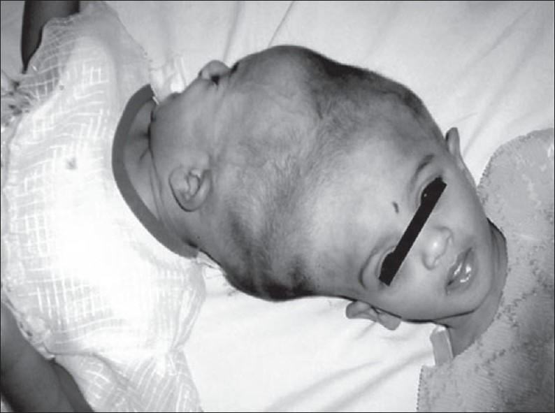
Craniopagus vertical type II twins
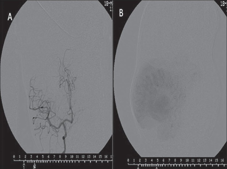
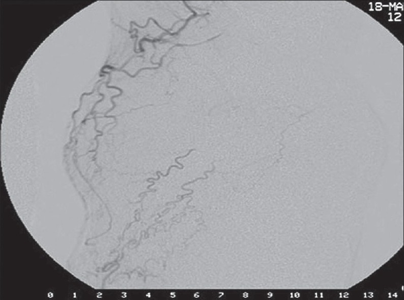
Internal carotid angiogram showing normal arterial phase(A) and capillary phase (B)of the twin I

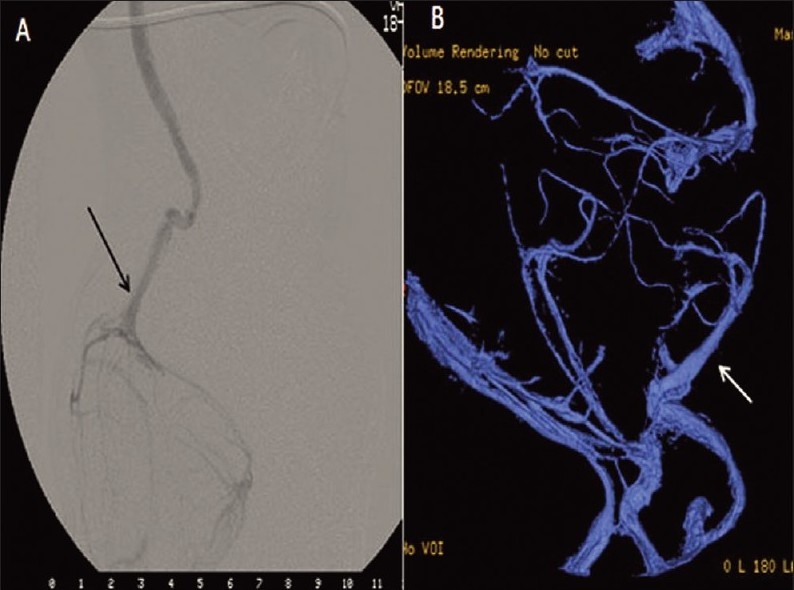
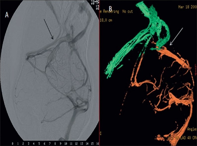
Showing the venous drainage of the twin I through the left transverse sigmoid system(arrow head) and circular sinus(arrow) into the twin II s transverse sigmoid jugular system bilaterally
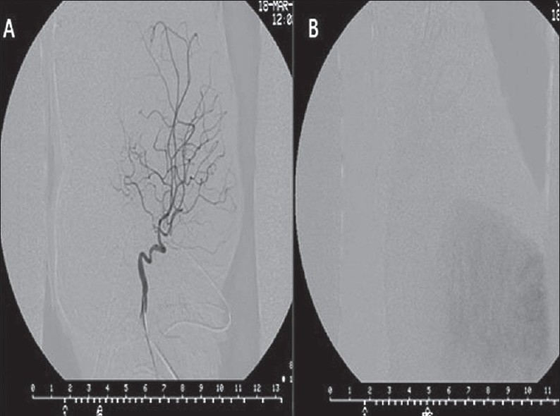
Internal carotid angiogram showing the normal arterial(A) and capillary phases(B) of the twin II
Venous drainage is through the circular sinus(arrow) into Transverse - Sigmoid -Jugular system bilaterally and the occipital sinus. Deep Venous system is not identifi ed clearly
External carotid angiogram of twin II shows signifi cant cross over of ECA territory to supply Twin I scalp
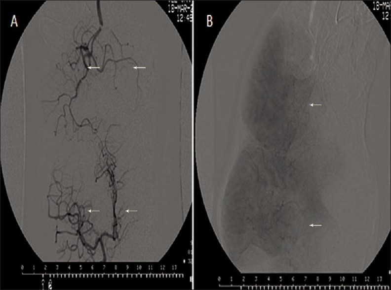
Simulataneous and sequential injections of both the twins showing normal arterial and capillary phases
Venous drainage is through the circular sinus into Transverse - Sigmoid -Jugular system bilaterally and the occipital sinus.A signifi cant portion of the venous drainage is shunted into Twin I s left transverse sinus. Deep Venous system is not identifi ed clearly
Similar articles
-
Preoperative Evaluation of Craniopagus Twins: Anatomy, Imaging Techniques, and Surgical Management.AJNR Am J Neuroradiol. 2020 Jun;41(6):951-959. doi: 10.3174/ajnr.A6571. Epub 2020 May 21. AJNR Am J Neuroradiol. 2020. PMID: 32439641 Free PMC article. Review.
-
Neurointerventional participation in craniopagus separation.Interv Neuroradiol. 2015 Aug;21(4):552-7. doi: 10.1177/1591019915590313. Epub 2015 Jun 10. Interv Neuroradiol. 2015. PMID: 26063696 Free PMC article.
-
Neuroradiological findings in adult cranially conjoined twins. Case report.J Neurosurg. 1998 Oct;89(4):635-9. doi: 10.3171/jns.1998.89.4.0635. J Neurosurg. 1998. PMID: 9761059
-
Separation of craniopagus twins in the era of modern neuroimaging, interventional neuroradiology, and frameless stereotaxy.Childs Nerv Syst. 2004 Aug;20(8-9):587-92. doi: 10.1007/s00381-004-0986-3. Epub 2004 Jul 27. Childs Nerv Syst. 2004. PMID: 15278383
-
A case of craniopagus parasiticus: an antenatal diagnosis by ultrasound screening at 16 weeks of gestation and a literature review of recently reported cases.Turk J Pediatr. 2019;61(6):941-945. doi: 10.24953/turkjped.2019.06.017. Turk J Pediatr. 2019. PMID: 32134590 Review.
Cited by
-
Preoperative Evaluation of Craniopagus Twins: Anatomy, Imaging Techniques, and Surgical Management.AJNR Am J Neuroradiol. 2020 Jun;41(6):951-959. doi: 10.3174/ajnr.A6571. Epub 2020 May 21. AJNR Am J Neuroradiol. 2020. PMID: 32439641 Free PMC article. Review.
-
Neurointerventional participation in craniopagus separation.Interv Neuroradiol. 2015 Aug;21(4):552-7. doi: 10.1177/1591019915590313. Epub 2015 Jun 10. Interv Neuroradiol. 2015. PMID: 26063696 Free PMC article.
References
-
- Hanson JW. Incidence of conjoined twinning. Lancet. 1975;2:1257. - PubMed
-
- Klucznik RP. Current technology and clinical applications of three-dimensional angiography. Radiol Clin North Am. 2002;40:711–28. - PubMed
-
- Schindler E, Hajek P. Craniopagus twins: Neuroradiological findings (CT, Angiography, MRI) Neuroradiolgy. 1988;30:11–6. - PubMed
-
- Kingston CA, McHugh K, Kumaradevan J, Kiely EM, Spitz L. Imaging in the preoperative assessment of conjoined twins. Radiographics. 2001;21:1187–208. - PubMed
-
- Barth RA, Filly RA, Goldberg JD, Moore P, Silverman NH. Conjoined twins: Prenatal diagnosis and assessment of associated malformations. Radiology. 1990;177:201–7. - PubMed
