. 2011;6(8):e23485.
doi: 10.1371/journal.pone.0023485. Epub 2011 Aug 24.

BPR1K653, a novel Aurora kinase inhibitor, exhibits potent anti-proliferative activity in MDR1 (P-gp170)-mediated multidrug-resistant cancer cells

Affiliations

BPR1K653, a novel Aurora kinase inhibitor, exhibits potent anti-proliferative activity in MDR1 (P-gp170)-mediated multidrug-resistant cancer cells

Chun Hei Antonio Cheung et al. PLoS One. 2011.

Abstract

Background: Over-expression of Aurora kinases promotes the tumorigenesis of cells. The aim of this study was to determine the preclinical profile of a novel pan-Aurora kinase inhibitor, BPR1K653, as a candidate for anti-cancer therapy. Since expression of the drug efflux pump, MDR1, reduces the effectiveness of various chemotherapeutic compounds in human cancers, this study also aimed to determine whether the potency of BPR1K653 could be affected by the expression of MDR1 in cancer cells.

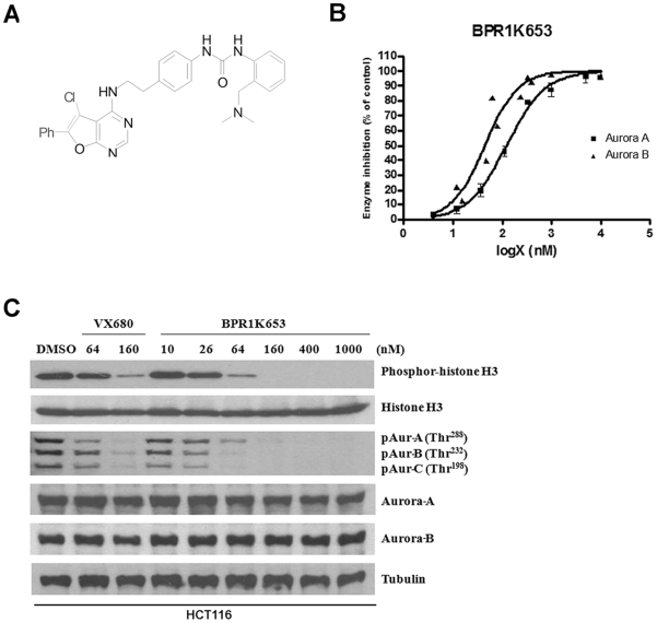
Principal findings: BPR1K653 specifically inhibited the activity of Aurora-A and Aurora-B kinase at low nano-molar concentrations in vitro. Anti-proliferative activity of BPR1K653 was evaluated in various human cancer cell lines. Results of the clonogenic assay showed that BPR1K653 was potent in targeting a variety of cancer cell lines regardless of the tissue origin, p53 status, or expression of MDR1. At the cellular level, BPR1K653 induced endo-replication and subsequent apoptosis in both MDR1-negative and MDR1-positive cancer cells. Importantly, it showed potent activity against the growth of xenograft tumors of the human cervical carcinoma KB and KB-derived MDR1-positive KB-VIN10 cells in nude mice. Finally, BPR1K653 also exhibited favorable pharmacokinetic properties in rats.

Conclusions and significance: BPR1K653 is a novel potent anti-cancer compound, and its potency is not affected by the expression of the multiple drug resistant protein, MDR1, in cancer cells. Therefore, BPR1K653 is a promising anti-cancer compound that has potential for the management of various malignancies, particularly for patients with MDR1-related drug resistance after prolonged chemotherapeutic treatments.


Conflict of interest statement

Competing Interests: The authors have declared that no competing interests exist.

Figures

Figure 1
Figure 1. BPR1K653 selectively inhibits the activity of Aurora kinases in vitro.
(A) Chemical structure of the anti-cancer compound BPR1K653. (B) BPR1K653 inhibited the activity of both Aurora-A and Aurora-B kinase as revealed by the in vitro kinase inhibition assay. (C) HCT116 cancer cells were treated with various concentrations of BPR1K653 and the commercially available pan-Aurora kinase inhibitor VX680, and the expression of various proteins were analyzed by Western blotting. Tubulin was used as the internal control.
Figure 2
Figure 2. Level of MDR1 expression correlates to the level of resistance of VX680/PHA739358 in KB-VIN10 and KB-S15 cancer cells.
Total mRNA was extracted from cells, and RT-PCR was performed to detect the expression of MDR1 in KB, KB-VIN10 and KB-S15 cells. GAPDH was used as internal control.
Figure 3
Figure 3. BPR1K653 induces endo-replication in both MDR1-negative and MDR1-expressing cancer cells.
(A, B and C) KB and KB-VIN10 cells were treated with BPR1K653 and VX680 for 48 h. (A) Nucleus were stained blue with Hoechst dye and microtubules were labeled red with the Alexa Fluor®-tagged anti-tubulin antibody. (B and C) Cells were incubated with propidium iodide and subsequently analyzed by flow cytometry. (D and E) HONE-1 cells were treated with BPR1K653 for 48 h. (D) Nucleus were stained blue with the Hoechst dye. (E) Cells were incubated with propidium iodide and subsequently analyzed by flow cytometry.
Figure 4
Figure 4. BPR1K653 down-regulates Histone H3 phosphorylation and cyclin B1 expression in both MDR1-negative and MDR1-expressing cancer cells.
(A) KB cells were treated with BPR1K653 and VX680 for 48 h and expression of various proteins were determined by Western blot analysis. Relative band intensities were shown. (B) KB-VIN10 cells were treated with either BPR1K653 or VX680 with/without verapamil for 48 h, and expression of various proteins was determined by Western blot analysis. Relative band intensities were shown. (C) HONE-1 cells were treated with BPR1K653 for 48 h, and expression of various proteins was determined by Western blot analysis. Actin was used as the internal control.
Figure 5
Figure 5. BPR1K653 induces apoptosis in both MDR1-negative and MDR1-expressing cancer cells.
(A, B and C) KB and KB-VIN10 cells were seeded on 8-well chamber slides overnight. (A) Cells were treated with either BPR1K653 or VX680 for 48 h. Translocation of the phosphatidylserine molecule in cells was analyzed by Annexin-V-FLUOS assay and cells were viewed using an UV-enabled microscope. General cell morphology was visualized by phase-contrast microscopy. (B) Cells were treated with either BPR1K653 or VX680 for 60 h and MagicRed™-DEVD Real-time Caspase-3/-7 Activity kit (Immunochemistry Technologies LLC) was used to detect the activation of caspase-3/-7 in cells, as indicated by the red fluorescent emission. Nucleus was counter-stained blue by Hoechst 33342, and cells were viewed real-time using an UV-enabled inverted microscope. (C and D) Detection of cells with DNA fragmentation by TUNEL assay. KB and KB-VIN10 cells were treated with either BPR1K653 or VX680 for 72 h. DNA fragmentations were analyzed using the TMR-red In Situ Cell Death Detection kit. Nucleus with DNA fragmentation was stained red. Nucleus was counter-stained blue by DAPI. Cells were analyzed by an UV-enabled microscope. (C) Representative photos were shown. (D) Labeled cells were counted, and percentage of apoptotic cells was calculated as follows: Total amount of the red fluorescent labeled (DNA fragmented) nucleus available ÷ Total amount of the blue fluorescent labeled nucleus available×100. Experiments were repeated twice. (E) BPR1K653 induces the cleavage of PARP in KB-VIN10 cancer cells. KB-VIN10 cells were treated with either BPR1K653 (2× IC50 of KB) or VX680 (2× IC50 of KB) with/without verapamil for 72 h. The cleavage of PARP was determined by Western blot analysis. Actin was used as the internal control.
Figure 6
Figure 6. Inhibition of human xenografts growth in vivo by BPR1K653.
(A, B, C and D) Nude mice bearing human cervical carcinoma KB xenografts were treated with vehicle control (⧫), 30 mg/kg VX680 for 5 days/week for 2 weeks (on days 6–10 and 13–17; ▴) or 15 mg/kg BPR0L075 for 5 days/week for 2 weeks (on days 6–10 and 13–17; •). (A) BPR1K653 treatment reduced the amount of the phosphor-Histone H3 positive cells present in tumor tissues. Immuno-histochemical analysis of the expression of phosphor-Histone H3 in the tumor tissue sections 24 h after the second BPR1K653 administration. Nucleus was stained blue/purple by hematoxylin and phosphor-Histone H3 was labeled in brown colour. Labeled cells were counted, and percentage of the phosphor-Histone H3 positive cells present in tumor tissues was calculated as follows: Total amount of cells with brown color labeled ÷ Total amount of cells available×100. Experiment was repeated twice. A statistically significant difference in the amount of phosphor-Histone H3 positive cells present in tumor tissues in mice treated with control versus BPR1K653 is denoted by “*”. *p<0.05. (B) Measurement of tumor volume. A statistically significant difference in tumor size in mice treated with control versus BPR1K653 and VX680 is denoted by “*”. *p<0.05. (C) Measurement of animal weight. (D) TUNEL analysis of the tumor tissue sections 12 days post-BPR1K653 treatment. Tumor tissue sections were analyzed by the FITC In Situ Cell death detection kit and fluorescent microscopy. Tissue treated with DNase was used as the positive control. Green fluorescence labeled nucleus indicates the induction of DNA fragmentation. Experiment was repeated twice. Quantitative analysis was shown. A statistically significant difference in the amount of apoptotic cells present in tumor tissues in mice treated with control versus BPR1K653 is denoted by “*”. *p<0.05. (E and F) Nude mice bearing the P-gp170/MDR-expressing KB-VIN10 xenograft was treated with vehicle control (⧫), 30 mg/kg VX680 for 5 days/week for 3 weeks (on days 12–16, 19–23 and 26–30; ▴) or 15 mg/kg BPR0L075 for 5 days/week for 3 weeks (on days 12–16, 19–23 and 26–30; •). (E) Measurement of tumor volume. A statistically significant difference in tumor size in mice treated with control versus BPR1K653 and VX680 is denoted by “*”. *p<0.05. (F) Measurement of animal weight. Data are the mean ± SD of tumor volume (mm3) at each time point (n = 5; *P<0.05).

Similar articles

Cited by

References

    1. Cheung CH, Coumar MS, Hsieh HP, Chang JY. Aurora kinase inhibitors in preclinical and clinical testing. Expert Opin Investig Drugs. 2009;18:379–398. - PubMed
    1. Cheung CH, Coumar MS, Chang JY, Hsieh HP. Aurora kinase inhibitor patents and agents in clinical testing: an update (2009–10) This article is an update to aurora kinase inhibitors review, which appeared in: Expert Opin. Ther. Patents 2009, 19, 1–36 and Expert Opin. Investig. Drugs 2009, 18, 1–20. Expert Opin Ther Pat. 2010;21:857–884. - PubMed
    1. Murata-Hori M, Tatsuka M, Wang YL. Probing the dynamics and functions of aurora B kinase in living cells during mitosis and cytokinesis. Mol Biol Cell. 2002;13:1099–1108. - PMC - PubMed
    1. Lu L-Y, Wood JL, Ye L, Minter-Dykhouse K, Saunders TL, et al. Aurora A is essential for early embryonic development and tumor suppression. Journal of Biological Chemistry. 2008;283:31785–31790. - PMC - PubMed
    1. Vischioni B, Oudejans JJ, Vos W, Rodriguez JA, Giaccone G. Frequent overexpression of aurora B kinase, a novel drug target, in non-small cell lung carcinoma patients. Mol Cancer Ther. 2006;5:2905–2913. - PubMed

Publication types

MeSH terms