. 2011;6(8):e23572.
doi: 10.1371/journal.pone.0023572. Epub 2011 Aug 24.

CK2 phosphorylation of Schistosoma mansoni HMGB1 protein regulates its cellular traffic and secretion but not its DNA transactions

Affiliations

CK2 phosphorylation of Schistosoma mansoni HMGB1 protein regulates its cellular traffic and secretion but not its DNA transactions

Isabel Caetano de Abreu da Silva et al. PLoS One. 2011.

Abstract

Background: The helminth Schistosoma mansoni parasite resides in mesenteric veins where fecundated female worms lay hundred of eggs daily. Some of the egg antigens are trapped in the liver and induce a vigorous granulomatous response. High Mobility Group Box 1 (HMGB1), a nuclear factor, can also be secreted and act as a cytokine. Schistosome HMGB1 (SmHMGB1) is secreted by the eggs and stimulate the production of key cytokines involved in the pathology of schistosomiasis. Thus, understanding the mechanism of SmHMGB1 release becomes mandatory. Here, we addressed the question of how the nuclear SmHMGB1 can reach the extracellular space.

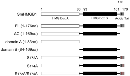
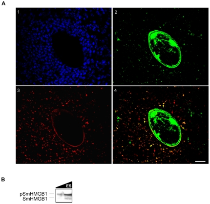
Principal findings: We showed in vitro and in vivo that CK2 phosphorylation was involved in the nucleocytoplasmic shuttling of SmHMGB1. By site-directed mutagenesis we mapped the two serine residues of SmHMGB1 that were phosphorylated by CK2. By DNA bending and supercoiling assays we showed that CK2 phosphorylation of SmHMGB1 had no effect in the DNA binding activities of the protein. We showed by electron microscopy, as well as by cell transfection and fluorescence microscopy that SmHMGB1 was present in the nucleus and cytoplasm of adult schistosomes and mammalian cells. In addition, we showed that treatments of the cells with either a phosphatase or a CK2 inhibitor were able to enhance or block, respectively, the cellular traffic of SmHMGB1. Importantly, we showed by confocal microscopy and biochemically that SmHMGB1 is significantly secreted by S. mansoni eggs of infected animals and that SmHMGB1 that were localized in the periovular schistosomotic granuloma were phosphorylated.

Conclusions: We showed that secretion of SmHMGB1 is regulated by phosphorylation. Moreover, our results suggest that egg-secreted SmHMGB1 may represent a new egg antigen. Therefore, the identification of drugs that specifically target phosphorylation of SmHMGB1 might block its secretion and interfere with the pathogenesis of schistosomiasis.


Conflict of interest statement

Competing Interests: The authors have declared that no competing interests exist.

Figures

Figure 1
Figure 1. Schematic diagram of the Schistosoma mansoni HMGB1 gene structure and constructed mutants.
SmHMGB1 full length (FL) consists of two DNA-binding domains, the HMG box A (aa 1–83), HMG box B (aa 84–169) and a short acidic C-terminal domain (170–176). ΔC (aa 1–169) refers to SmHMGB1 lacking only its acidic C-terminal domain; domain A (aa 1–83) refers to SmHMGB1 lacking its HMG box B domain; domain B (aa 84–169) refers to SmHMGB1 lacking its HMG box A domain; S172A refers to SmHMGB1 with a point mutation at serine 172, substituted by alanine; S174A refers to SmHMGB1 with a point mutation at serine 174, substituted by alanine; S172A/S174A refers to SmHMGB1 with two point mutations at serines 172 and 174, both substituted by alanine.
Figure 2
Figure 2. In vitro kinase assay of SmHMGB1 phosphorylation.
(A) One µg of the recombinant SmHMGB1 full length protein (FL) was used as a substrate for commercial CK2, at various incubation times, in the presence of [γ 32P]ATP. Heparin was included to show specific inhibition of CK2. Phosphorylations were analyzed by 15% SDS-PAGE and autoradiography (top panel). Bottom panel is the Coomassie blue stained gel of the SmHMGB1-FL used in the reactions; (B) S. mansoni adult worm total protein extract (4 µg) was used, as a source of kinases, in in vitro phosphorylation reactions. One µg of recombinant SmHMGB1-FL was incubated with the extract for 1 h in the presence of [γ 32P]ATP, and with (1.8 and 3.6 µM) or without TBBt, a specific CK2 inhibitor. TBBt was dissolved in DMSO and we used it as control. Top panel: phosphorylation; bottom panel: Coomassie blue staining; (C) One µg of recombinant SmHMGB1 full length protein (FL) was used as a substrate for commercial PKA and PKC in phosphorylation reactions for 1 h in the presence of γ[32P]ATP. These experiments were repeated three times.
Figure 3
Figure 3. Mapping of CK2 phosphorylation sites in SmHMGB1.
(A) One µg of the recombinant SmHMGB1-FL, SmHMGB1-ΔC, SmHMGB1-domain A and SmHMGB1-domain B were assayed for CK2 phosphorylation, as described in Figure 2. Top panel shows phosphorylation of SmHMGB1-FL (lane 1); no phosphoprylation was observed with the deleted constructions (lanes 2–4). Bottom panel shows the Coomassie blue staining of the purified recombinant proteins used in the phosphorylation assay. (B) Only part of the protein is represented (from aa 162 to the end of the protein) to show where the point mutations took place. The two serine residues located at the acidic C-terminal tail of SmHMGB1-FL were substituted to alanines (in red), accordingly. (C) One µg of the recombinant SmHMGB1-FL, SmHMGB1-ΔC or mutated constructs were assayed for CK2 phosphorylation. These experiments were repeated three times.
Figure 4
Figure 4. DNA supercoiling and bending assays by phosphorylated SmHMGB1.
(A) Circular relaxed plasmid pTZ19R DNA was incubated in the presence of topoisomerase I with 1 µg of recombinant SmHMGB1-FL or SmHMGB1-S172A/S174A that were phosphorylated (lanes 3–5) or not (lanes 6–8 and 9–11), by CK2. Deproteinized DNA topoisomers were resolved on 1% agarose gels, followed by staining of the gels with ethidium bromide. Form I, supercoiled DNA; form II, relaxed circular DNA. (B) Top panel: autoradiography; bottom panel: Coomassie staining. (C) A 32P-labeled 123-bp DNA fragment (∼1 nM) was pre-incubated with 50 ng of recombinant proteins, that were phosphorylated (lanes 7–9) or not (lanes 4–6, 10–12, 13–15 and 16–18), followed by ligation with T4 DNA ligase. Exonuclease III was used to verify the identity of DNA circles. The deproteinized DNA ligation products were subjected to electrophoresis on 6% non-denaturing polyacrylamide gels and visualized by autoradiography. Controls are as follows: FL(c1): SmHMGB1-FL without CK2; FL(c2): SmHMGB1-FL without phosphate; FL(c3): SmHMGB1-FL without CK2 buffer. Linear: linear DNA; Lm: linear multimers. (D) Top panel: autoradiography; bottom panel: Coomassie staining. These experiments were repeated four times.
Figure 5
Figure 5. Phosphorylation of SmHMGB1 mediates its cellular traffic in HeLa cells.
HeLa cells were transfected with empty control plasmid pEGFP, pEGFP-SmHMGB1-FL or pEGFP-SmHMGB1-S172A/S174A plasmids and untreated (panels A) or treated with 100 nM okadaic acid (OA) for 6 h (panels B) or with OA+75 µM TBBt (panels C). SmHMGB1-EGFP fusion proteins were detected by fluorescence microscopy. Nuclei were stained with DAPI. Cell viability was assessed by Trypan blue and LDH activity (data not shown). Scale bar 3 µm. This result is a representative of four independent experiments.
Figure 6
Figure 6. In situ localization of native SmHMGB1 protein in the nucleus and cytoplasm of S. mansoni cells.
(a and b) Transmission electron microscopy (TEM) of cells from S. mansoni male adult worms showing the nucleus (N), nucleolus (Nc), cytoplasm (C) and the nuclear membrane (arrowheads). Insets a1, a2, b1 and b2 depict a closer visualization of the interface between the nucleus and the cytoplasm. The immunogold staining shows the labeling of SmHMGB1 in both cellular compartments (arrows indicate representative SmHMGB1 labeling). Bars: a and b = 100 nm; a1 and a2 = 50 nm. This image is a representative of several cells observed under TEM.
Figure 7
Figure 7. Endogenous phosphorylation of SmHMGB1 did not alter its DNA bending activity.
(A) Western blot analysis was carried out with S. mansoni protein extracts and an anti-SmHMGB1 polyclonal antibody. SmHMGB1 antibodies detected two proteins in the total extract (top panel, lane 1). The lower molecular weight protein was detected in the nuclear extract only (top panel, lane 2); the higher molecular weight protein was detected in the cytosolic extract only (top panel, lane 3). SmHMGB1 that was immunopreciptated from the nuclear or cytosolic extracts using SmHMGB1 antibodies were reacted against anti-phosphoserine antibodies in a Western blot (bottom panel, lane 2 (nuclear extract) and 3 (cytosolic extract); phosphorylation of SmHMGB1 at serines is indicated by pSmHMGB1). (B) Bending assay: a 32P-labeled 123-bp DNA fragment (1 nM) was pre-incubated with 10 µg of total (lane 4), 4 µg of nuclear (lane 5) or 4 µg of cytosolic (lane 6) protein extracts from S. mansoni adult worms, and the assay performed as described in Figure 4C. To make sure that the circles were formed by the activity of SmHMGB1, nuclear or cytosolic (lanes 7 and 8, respectively) extracts were incubated with anti-SmHMGB1 antibodies prior bending reactions. Extracts that were incubated with SmHMGB1 antibody did not show any circularization (or bending) activity (lanes 7 and 8). The Exo III control proved the identity of the circles. (C) Top panel: SDS-PAGE of S. mansoni adult worm nuclear (N) or cytosolic (C) extracts. Bottom panel: Western blot with anti-acetylated histones, showing no cross-contamination between the two extracts. These experiments were repeated three times.
Figure 8
Figure 8. Localization of phosphorylated SmHMGB1 in the granulomatous liver.
(A) Immunostaining of hepatic granuloma with a S. mansoni egg in the center. Nuclei were stained with DAPI (1); Detection of SmHMGB1 using an anti-SmHMGB1 polyclonal antibody (2). Detection of phosphorylated SmHMGB1 using an anti-phosphoserine monoclonal antibody (3). A significant amount of secreted SmHMGB1 detected in the granulomatous liver is phosphorylated (merged images of panels 2 and 3). In panel 4, green arrows point to secreted but non-phosphorylated SmHMGB1; red arrows point to phosphorylated proteins from the host; orange arrows point to secreted phosphorylated SmHMGB1. Controls with the pre-immune sera (not shown) or with the secondary antibody only (Figure S3), exhibited a residual auto-fluorescence from the eggshell. Scale bar: 20 µm. This figure is a representative of the several egg-induced granuloma analyzed from three independent mice livers. (B) Eggs (∼106 eggs) from these livers were processed and egg secretions (ES) were assayed by Western blot using anti-SmHMGB1 antibody. The two isoforms (phosphorylated and unphosphorylated) of SmHMGB1 were detected in egg secretions. However, the high molecular weight (phosphorylated) isoform is significantly more abundant in egg secretions (top band).

Similar articles

Cited by

References

    1. WHO. TDR Strategic Direction for Research. Schistosomiasis World Health Organization. Geneve; 2002.
    1. Oliveira VR, El-Cheikh MC, Aguiar AM, Balduino A, de Fatima BPM, et al. Schistosoma mansoni egg-induced hepatic granulomas in mice deficient for the interferon-gamma receptor have altered populations of macrophages, lymphocytes and connective tissue cells. Microbes Infect. 2000;2:1817–1826. - PubMed
    1. Boros DL. Immunoregulation of granuloma formation in murine schistosomiasis mansoni. Ann N Y Acad Sci. 1986;465:313–323. - PubMed
    1. Caldas IR, Campi-Azevedo AC, Oliveira LF, Silveira AM, Oliveira RC, et al. Human schistosomiasis mansoni: immune responses during acute and chronic phases of the infection. Acta Trop. 2008;108:109–117. - PubMed
    1. Stros M. HMGB proteins: interactions with DNA and chromatin. Biochim Biophys Acta. 2010;1799:101–113. - PubMed

Publication types

MeSH terms