. 2011;6(8):e23577.
doi: 10.1371/journal.pone.0023577. Epub 2011 Aug 24.

Serum-nutrient starvation induces cell death mediated by Bax and Puma that is counteracted by p21 and unmasked by Bcl-x(L) inhibition

Affiliations

Serum-nutrient starvation induces cell death mediated by Bax and Puma that is counteracted by p21 and unmasked by Bcl-x(L) inhibition

Frédérique Braun et al. PLoS One. 2011.

Abstract

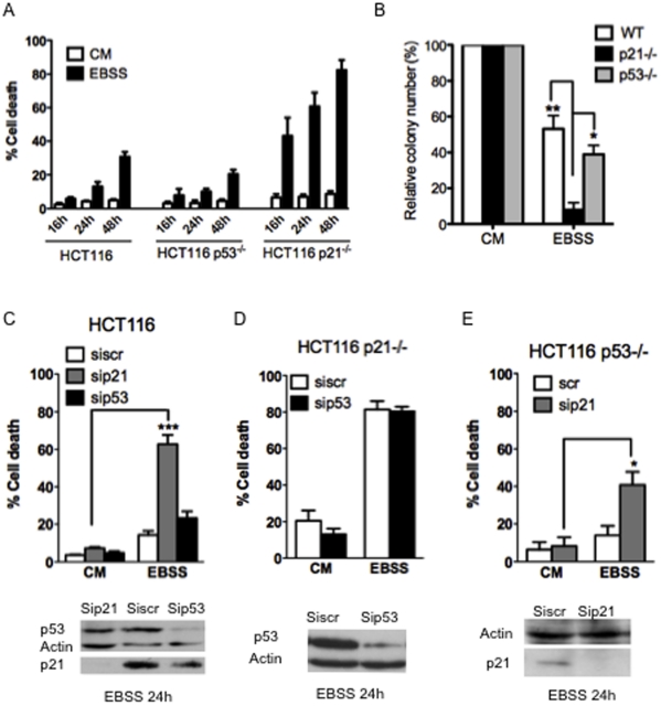
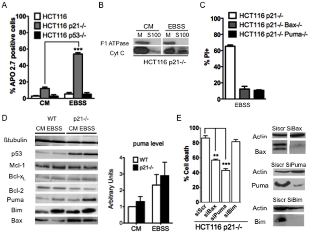
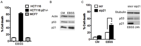
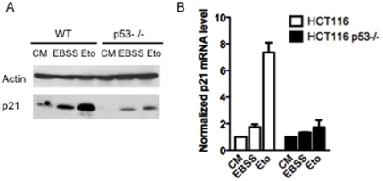
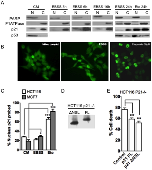
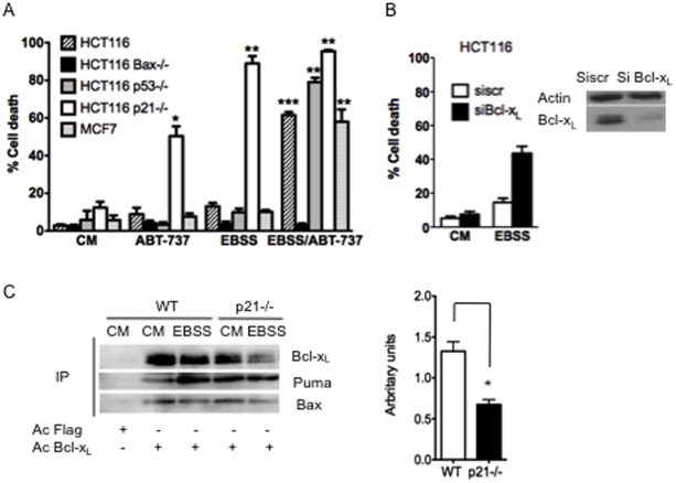
The cyclin-dependent kinase inhibitor p21 (p21WAF1/Cip1) is a multifunctional protein known to promote cell cycle arrest and survival in response to p53-dependent and p53 independent stimuli. We herein investigated whether and how it might contribute to the survival of cancer cells that are in low-nutrient conditions during tumour growth, by culturing isogenic human colorectal cancer cell lines (HCT116) and breast cancer cell lines in a medium deprived in amino acids and serum. We show that such starvation enhances, independently from p53, the expression of p21 and that of the pro-apoptotic BH3-only protein Puma. Under these conditions, p21 prevents Puma and its downstream effector Bax from triggering the mitochondrial apoptotic pathway. This anti-apoptotic effect is exerted from the cytosol but it is unrelated to the ability of p21 to interfere with the effector caspase 3. The survival function of p21 is, however, overcome by RNA interference mediated Bcl-x(L) depletion, or by the pharmacological inhibitor ABT-737. Thus, an insufficient supply in nutrients may not have an overt effect on cancer cell viability due to p21 induction, but it primes these cells to die, and sensitizes them to the deleterious effects of Bcl-x(L) inhibitors regardless of their p53 status.


Conflict of interest statement

Competing Interests: The authors have declared that no competing interests exist.

Figures

Figure 1
Figure 1. Role of p21 and p53 in cell death induced by serum nutrient starvation.
(A) The indicated cells were placed in complete medium (CM) or in starved medium for 16 h, 24 h or 48 h (EBSS) and cell death was assayed by a trypan blue-staining. Data presented are mean ± SEM of three independent experiments. (B) The indicated cells were placed in complete medium (CM) or in starved medium during 24 h and then seeded at a low density for 2 weeks in complete medium before visualization of viable clones by crystal violet staining. (C, D and E) The indicated HCT116 cells were transfected with the indicated siRNA. 48 h later, cells were starved in EBSS medium for 24 h (EBSS) or not (CM). Western blot analysis was performed to confirm silencing and cell death was assayed by a trypan blue-staining. Data presented are mean ± SEM of three independent experiments.
Figure 2
Figure 2. Puma and Bax as key actors of apoptosis induced by starvation in p21-negative HCT116 cells.
(A) The indicated cells were placed in complete medium (CM) or in starved medium for 24 h (EBSS). Apoptosis was assayed by APO 2.7 staining using flow cytometry. Data are expressed as a percentage of the total population. (B) Mitochondria (M) and S100 (S100) lysates from HCT116 p21−/− cells, starved 16 h in EBSS medium (EBSS) or not (CM), were analysed by western blot analysis for F1ATPase and Cytochrome C (Cyt C). (C) Viability of the indicated cells placed under starvation during 24 h was analyzed by PI staining followed by flow cytometry. (D) The indicated cells were placed under complete medium (CM) or in starved medium (EBSS). After 24 h, protein extracts were prepared and the expression of the indicated protein was analysed by western blotting. The amount of Puma was evaluated in each condition by densitometric analysis from three independent experiments and normalized to the amount of Puma level measured in complete medium in wild-type cells (E) The p21 negative HCT116 cells were transfected with the indicated siRNA. 48 h later, cells were starved in EBSS medium for 24 h. Western blot analysis of Bax, Puma and Bim was performed and cell death was assayed by a trypan blue-staining. Data presented in (A), (C) and (E) are mean ± SEM of three independent experiments.
Figure 3
Figure 3. Survival function of p21 in the Caspase 3 deficient MCF7 cells.
(A) The indicated cells were starved in EBSS medium for 24 h (EBSS) and cell death was assayed by a trypan blue-staining. (B) MCF7 cells were placed in complete medium (CM) or starved in EBSS medium (EBSS) for 24 h. Western blot analysis was performed to analyse p21 and p53 expression. (C) MCF7 cells were transfected with the indicated siRNA. 48 h later, cells were starved in EBSS medium for 24 h (EBSS) or not (CM). Western blot analysis of p21 and p53 was performed and cell death was assayed by a trypan blue-staining. Data presented in (A) and (C) are mean ± SEM of three independent experiments.
Figure 4
Figure 4. Serum-nutrient starvation enhancement of p21 protein levels.
(A) The indicated cells were placed in complete medium (CM), treated 24 h with 50 µM of etoposide (Eto) or starved 24 h in EBSS medium (EBSS). Western analysis was performed to detect p21. (B) The indicated cells were placed in complete medium (CM), treated with 50 µM of etoposide (Eto) or starved in EBSS medium (EBSS) during 6 h for qPCR analysis. p21 mRNA level is expressed as relative values with two house-keeping genes and normalized to complete medium condition.
Figure 5
Figure 5. Impact of intracellular localization on the survival function of p21.
(A) Nuclear versus cytoplasmic extracts prepared at the indicated times from HCT116 wt cells place under EBSS or not (CM), or treated with 50 µM of etoposide (Eto) for 24 h. Western analysis was performed for p21, p53 and F1ATPase and PARP. (B) Immunostaining of p21 in HCT116 wt cells placed under complete medium (CM), starved medium (EBSS) or treated with 50 µM of etoposide (Eto) for 24 h. Nucleus was stained with DAPI. (C) Quantification of immunostaining of p21 in HCT116 and MCF7 cells. The amount of cells containing a nuclear p21 localisation was expressed as a percentage of total cells detected with DAPI staining. (D) Western blot analysis of ectopic p21 in HCT116 p21−/− transfected with either pGFPires21FL expressing the full-length p21 protein (FL) or with pGFPires21ΔNSL (ΔNSL) expressing the p21 protein deleted from its C-terminal NSL domain. (E) HCT116 p21−/−transfected cells with the indicated vector were placed in EBSS medium and followed by time-lapse videomicroscopy. The vectors used allowed the expression of the GFP concomitant with p21, due to the presence of an IRES element. The percentage of transfected (ie GFP-expressing) cells exhibiting morphological features of cell death was assayed during 24 h. Data presented in (C) and (E) are mean ± SEM of three independent experiments.
Figure 6
Figure 6. Sensitization to starvation induced cell death by down regulation or inhibition of Bcl-xL.
(A) Inhibition of the BH3 domain of Bcl-2/Bcl-xL sensitizes starved cells independently of p53. The indicated cells were placed in complete medium (CM) or starved in EBSS medium for 24 h (EBSS) and treated with 1 µM of ABT-737 or not and cell death determined by blue trypan assay (**, ***, significantly different from cells placed in complete medium). (B) HTC116 cells were transfected with the indicated siRNA. 48 h later, cells were starved in EBSS medium for 24 h (EBSS) or not (CM). Western analysis of Bcl-xL was performed and cell death assayed by a trypan blue-staining. (C) Wild-type and p21−/− HCT116 cells were placed in complete medium (CM) or starved in EBSS medium for 24 h (EBSS). Expression of Bcl-xL and Puma and Bax in cell lysates was analysed by western blot. Cells lysates were immunoprecipated (IP) with an anti-Bcl-xL antibody or an anti-Flag antibody as a control, and the presence of Bcl-xL and Puma and Bax in the immunoprecipitated fractions was analysed by immunoblotting. The amount of Puma that co-immunoprecipitated with Bcl-xL (evaluated by densitometric analysis and normalized to the amount of immunoprecipitated Bcl-xL) in EBSS condition divided by the amount of Puma that co-immunoprecipitated with Bcl-xL (evaluated by densitometric analysis and normalized to the amount of immunoprecipitated Bcl-xL) in complete medium condition are shown in wilt type and p21 deficient cells as indicated. Data presented in A, B and C are mean ± SEM of three independent experiments.

References

    1. Gartel AL, Tyner AL. The role of the cyclin-dependent kinase inhibitor p21 in apoptosis. Mol Cancer Ther. 2002;1:639–49. - PubMed
    1. el-Deiry WS, Tokino T, Velculescu VE, Levy DB, Parsons R, et al. WAF1, a potential mediator of p53 tumor suppression. Cell. 1993;75:817–25. - PubMed
    1. Coqueret O. New roles for p21 and p27 cell-cycle inhibitors: a function for each cell compartment? Trends Cell Biol. 2003;13:65–70. - PubMed
    1. Jung YS, Qian Y, Chen X. Examination of the expanding pathways for the regulation of p21 expression and activity. Cell Signal. 2010;22:1003–12. - PMC - PubMed
    1. Janicke RU, Sohn D, Essmann F, Schulze-Osthoff K. The multiple battles fought by anti-apoptotic p21. Cell Cycle. 2007;6:407–13. - PubMed

Publication types

MeSH terms