. 2011;6(8):e23643.
doi: 10.1371/journal.pone.0023643. Epub 2011 Aug 24.

Rectification of the water permeability in COS-7 cells at 22, 10 and 0°C

Affiliations

Rectification of the water permeability in COS-7 cells at 22, 10 and 0°C

Diana B Peckys et al. PLoS One. 2011.

Abstract

The osmotic and permeability parameters of a cell membrane are essential physico-chemical properties of a cell and particularly important with respect to cell volume changes and the regulation thereof. Here, we report the hydraulic conductivity, L(p), the non-osmotic volume, V(b), and the Arrhenius activation energy, E(a), of mammalian COS-7 cells. The ratio of V(b) to the isotonic cell volume, V(c iso), was 0.29. E(a), the activation energy required for the permeation of water through the cell membrane, was 10,700, and 12,000 cal/mol under hyper- and hypotonic conditions, respectively. Average values for L(p) were calculated from swell/shrink curves by using an integrated equation for L(p). The curves represented the volume changes of 358 individually measured cells, placed into solutions of nonpermeating solutes of 157 or 602 mOsm/kg (at 0, 10 or 22°C) and imaged over time. L(p) estimates for all six combinations of osmolality and temperature were calculated, resulting in values of 0.11, 0.21, and 0.10 µm/min/atm for exosmotic flow and 0.79, 1.73 and 1.87 µm/min/atm for endosmotic flow (at 0, 10 and 22°C, respectively). The unexpected finding of several fold higher L(p) values for endosmotic flow indicates highly asymmetric membrane permeability for water in COS-7. This phenomenon is known as rectification and has mainly been reported for plant cell, but only rarely for animal cells. Although the mechanism underlying the strong rectification found in COS-7 cells is yet unknown, it is a phenomenon of biological interest and has important practical consequences, for instance, in the development of optimal cryopreservation.


Conflict of interest statement
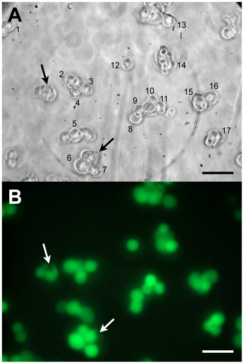
Competing Interests: The authors have declared that no competing interests exist.

Figures

Figure 1
Figure 1. COS-7 cells suspended in the MicroCell chamber at 22°C.
A) First image from a series of images initiated 21 s after cells had been exposed to hypotonic (157 mOsm/kg) TBS. The numbers indicate individually identified cells selected for analysis. The changes in their diameters were recorded until well after the cells had reached their maximal volume. Arrows point to two cells that show membrane damage in the form of a bleb of leaking cytosol. B) Fluorescence image of the same group of cells taken immediately after the last image of the bright light image series, about 5 min after A) was taken. Viable cells display green calcein AM fluorescence. Although the two damaged cells still display fluorescence, such cells were excluded from the analysis. Scale bar = 50 µm.
Figure 2
Figure 2. Boyle-van't Hoff plot of COS-7 cells.
The BvH plot displays the absolute volume as a function of the reciprocal of the osmolality of the TBS in which the cells were suspended for 5 to 7 min at 22°C. Each point is the mean of 250 to 828 individually analyzed cells. The volumes were calculated from the measured diameters as described in the text. Data were fitted by the method of least squares.
Figure 3
Figure 3. Normalized shrink curves of COS-7 cells in hypertonic (602 mOsm/kg) TBS.
The cell volumes were normalized to the mean isotonic volume at each temperature; namely, 3324, 3920, and 3853 µm3. The data represent averages and S.E. values of 153 individually measured cells. Cells reached their equilibrium volume faster the higher the temperatures.
Figure 4
Figure 4. Normalized swell curves of COS-7 cells in hypotonic (157 mOsm/kg) TBS.
The data represent averages and S.E. values of 205 individually measured cells. Cells reached their equilibrium volume faster the higher the temperatures. However, these times were significantly longer than in the hyperosomotic situation for all three temperatures tested.
Figure 5
Figure 5. Lp values (water permeability) obtained under hyper- and hypotonic conditions.
The three bars to the left show the Lp's under hypertonic conditions and the three bars to the right under hypotonic conditions. The differences between the two Lp's determined at the same temperature are highly significant (p<0.0001). That is true for all three temperatures. These differences strongly indicate rectification of the water flow; that is, the flow rate is different out of the cell than into the cell.
Figure 6
Figure 6. Arrhenius plots for COS-7 cells.
The upper line represents the fit for hypotonic conditions (157 mOsm/kg), indicated by circles, and the lower line is the fit for hypertonic conditions (602 mOsm/kg), indicated by triangles, at temperatures of 0 and 10°C. The activation energy, Ea, of the hypotonic Lp values was 12.0 kcal/mol. The value for Ea of the hypertonic Lp values was 10.7 kcal/mol. The Lp values gained at 22°C (open symbols) were not used for the calculation of Ea, for reasons given in the discussion.
Figure 7
Figure 7. Comparison of the experimental data with computer simulations.
Plots of the volumes of COS-7 cells as function of time under hypotonic conditions (left column) and under hypertonic conditions (right column). The diamonds are the experimental values. The light solid line is the simulated curve obtained by calculating the cell volumes as a function of time using the classical differential equation [Eq 3]. The heavy solid line was obtained by sliding the light line in the X and Y directions to obtain the best fit by eye to the experimental points.

References

    1. Mazur P. Kinetics of Water Loss from Cells at Subzero Temperatures and the Likelihood of Intracellular Freezing. J Gen Physiol. 1963;47:347–369. - PMC - PubMed
    1. Mazur P. Principles of Cryobiology. In: Fuller NLBJ, Benson EE, editors. Life in the Frozen State. Boca Raton: CRC Press; 2004. pp. 3–65.
    1. Mazur P, Seki S, Pinn IL, Kleinhans FW, Edashige K. Extra- and intracellular ice formation in mouse oocytes. Cryobiology. 2005;51:29–53. - PubMed
    1. Seki S, Mazur P. The temperature and type of intracellular ice formation in preimplantation mouse embryos as a function of the developmental stage. Biol Reprod. 2010;82:1198–1205. - PMC - PubMed
    1. Kleinhans FW, Guenther JF, Roberts DM, Mazur P. Analysis of intracellular ice nucleation in Xenopus oocytes by differential scanning calorimetry. Cryobiology. 2006;52:128–138. - PubMed

Publication types