. 2011;6(8):e23763.
doi: 10.1371/journal.pone.0023763. Epub 2011 Aug 24.

Ischemic tolerance protects the rat retina from glaucomatous damage

Affiliations

Ischemic tolerance protects the rat retina from glaucomatous damage

Nicolás Belforte et al. PLoS One. 2011.

Abstract

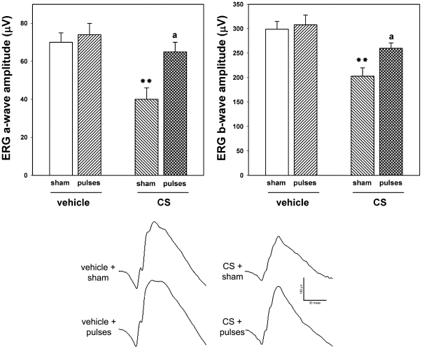
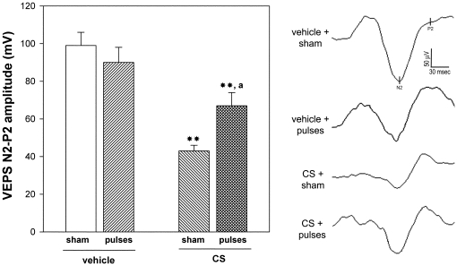
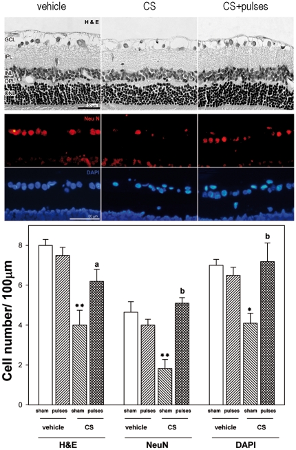
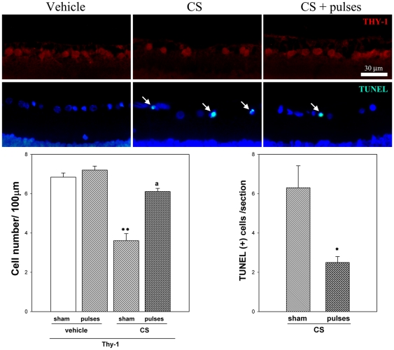
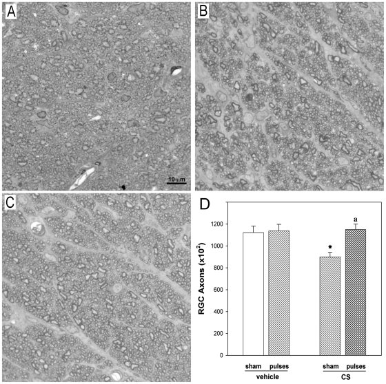
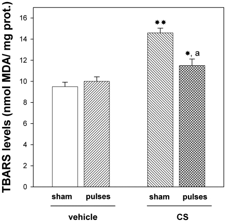
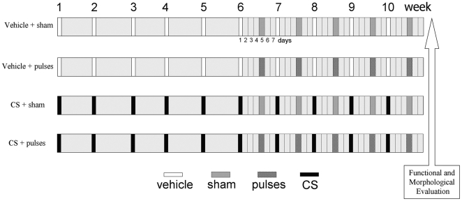
Glaucoma is a leading cause of acquired blindness which may involve an ischemic-like insult to retinal ganglion cells and optic nerve head. We investigated the effect of a weekly application of brief ischemia pulses (ischemic conditioning) on the rat retinal damage induced by experimental glaucoma. Glaucoma was induced by weekly injections of chondroitin sulfate (CS) in the rat eye anterior chamber. Retinal ischemia was induced by increasing intraocular pressure to 120 mmHg for 5 min; this maneuver started after 6 weekly injections of vehicle or CS and was weekly repeated in one eye, while the contralateral eye was submitted to a sham procedure. Glaucoma was evaluated in terms of: i) intraocular pressure (IOP), ii) retinal function (electroretinogram (ERG)), iii) visual pathway function (visual evoked potentials, (VEPs)) iv) histology of the retina and optic nerve head. Retinal thiobarbituric acid substances levels were assessed as an index of lipid peroxidation. Ischemic conditioning significantly preserved ERG, VEPs, as well as retinal and optic nerve head structure from glaucomatous damage, without changes in IOP. Moreover, ischemia pulses abrogated the increase in lipid peroxidation induced by experimental glaucoma. These results indicate that induction of ischemic tolerance could constitute a fertile avenue for the development of new therapeutic strategies in glaucoma treatment.


Conflict of interest statement

Competing Interests: The authors have declared that no competing interests exist.

Figures

Figure 1
Figure 1. Electroretinographic preservation in hypertensive eyes induced by the application of brief ischemia pulses.
ERGs were registered after 10 weeks of treatment with vehicle or CS. CS induced a significant decrease in ERG a- and b-wave amplitude, as compared with vehicle-injected eyes. In hypertensive eyes submitted to ischemia pulses, a significant reversion of these alterations was observed. The lower panel shows representative scotopic ERG traces from eyes injected with vehicle or CS without or with ischemia pulses. Data are the mean ± SEM (n = 10 animals per group); **p<0.01 versus vehicle injected eyes without ischemia pulses (sham); a: p<0.05, versus CS-injected eyes without ischemia pulses (sham), by Tukey's test.
Figure 2
Figure 2. Flash VEPs in eyes injected with vehicle or CS without or with ischemia pulses.
Animals were weekly injected with vehicle or CS for 10 weeks. Ischemia pulses were applied in one eye, while the contralateral eye was submitted to a sham procedure. Left panel shows average amplitudes of VEP N2-P2 component amplitude and right panel shows representative VEP traces. A significant reduction in flash VEP N2-P2 amplitude component was observed in eyes injected with CS for 10 weeks without ischemia pulses. The application of weekly ischemia pulses significantly abrogated the effect of ocular hypertension. No changes between vehicle injected eyes without or with ischemia pulses were observed. Data are mean ± SEM (n = 10 eyes/group), **p<0.01 versus vehicle injected eyes without ischemia pulses (sham), a: p<0.05 versus CS-injected eyes without ischemia pulses (sham), by Tukey's test.
Figure 3
Figure 3. Retinal histology examination after 10 weeks of ocular hypertension.
Upper panel: Representative photomicrographs of retinal sections stained with hematoxylin and eosin from a vehicle-injected eye, and a hypertensive eye without or with pulses of ischemia. Note the diminution of GCL cells in the eye injected with CS without ischemia pulses. The application of ischemia pulses preserved this parameter. The other retinal layers showed a normal appearance in all groups. Middle panel: Immunohistochemical detection of NeuN-positive neurons in the GCL from a vehicle-injected eye, a hypertensive eye without or with ischemia pulses. A strong NeuN-immunostaining (red) was confined to ganglion cells in the GCL. The number of NeuN positive ganglion cells was lower in hypertensive eyes without ischemia pulses than in vehicle- injected eyes, whereas the application of ischemia pulses in CS-injected eyes increased NeuN-immunostaining. A similar profile was observed for cell nuclei counterstained with DAPI (blue). Lower Panel: cell count in the GCL evaluated by H&E staining, NeuN immunostaining, and DAPI labeling. By all these methods, a significant decrease of the number of cells in the GCL was observed in CS- injected eyes without ischemia pulses as compared with vehicle-injected eyes (sham), whereas ischemia pulses significantly preserved this parameter in CS-injected eyes. Scale bar: Upper panel  =  50 µm; Middle panel  =  50 µm. Data are the mean ± SEM (n = 5 animals per group). * p<0.05, ** p<0.01 vehicle injected eyes without ischemia pulses (sham), a: p<0.05, b: p<0.01 versus CS-injected eyes without ischemia pulses (sham), by Tukey's test. GCL, ganglion cell layer; IPL, inner plexiform layer; INL, inner nuclear layer; OPL, outer plexiform layer; ONL, outer nuclear layer.
Figure 4
Figure 4. Thy-1 level and TUNEL assessment after 10 weeks of ocular hypertension.
Representative photomicrographs showing Thy-1 immunofluorescence (upper panel) and TUNEL analysis (middle panel) from a vehicle-injected eye, and a hypertensive eye without or with pulses of ischemia. Note TUNEL+ cells in the GCL (arrows). Cell nuclei were counterstained with DAPI. Lower panel: The number of Thy-1 positive cells was significantly lower in hypertensive eyes without ischemia pulses than in vehicle- injected eyes, whereas the application of ischemia pulses in CS-injected eyes increased Thy-1 immunostaining. The number of TUNEL+ cells in the GCL was significantly higher in hypertensive eyes submitted to a sham procedure than in those submitted to ischemia pulses. No TUNEL+ cells were observed in vehicle-injected eyes submitted to ischemia pulses or sham procedure (not shown). Scale bar: 30 µm. Data are mean ± SEM (n = 5 eyes per group). For TUNEL analysis: * p<0.05 versus CS-injected eyes submitted to sham procedure, by Students t-test; For Thy-1 analysis: **p<0.01 versus vehicle injected eyes without ischemia pulses (sham), a: p<0.01, versus CS-injected eyes without ischemia pulses (sham), by Tukey's test.
Figure 5
Figure 5. ONH sections from a control or a CS-treated eye without or with ischemia pulses.
(A) Healthy, intact control optic nerve. Note the homogeneity of the staining. In vehicle-injected eyes, individual axons were generally uniform in shape, rounded and packed together tightly to form the fibers of the healthy nerve. In CS-treated eye without ischemia pulses (B) a less stained area indicates a nerve alteration. Disease in individual axons was characterized by axonal distention and distortion that resulted in a departure from the circular morphology of normal axons. In contrast, a conserved structure of the ONH was observed in the CS-treated eye with ischemia pulses (C). Toluidine blue. (D) Number of axons in eyes injected with vehicle or CS without or with ischemia pulses. A significant decrease in the axon number was observed in CS- injected eyes without ischemia pulses as compared with vehicle-injected eyes (sham), whereas ischemia pulses significantly preserved this parameter. Scale bar: 10 µm. Data are mean ± SEM (n = 5 eyes/group) *p<0.05 vehicle injected eyes without ischemia pulses (sham), a: p<0.05 versus CS-injected eyes without ischemia pulses (sham), by Tukey's test.
Figure 6
Figure 6. Retinal TBARS levels in animals injected with CS or vehicle, without o with ischemia pulses.
This parameter was significantly higher in eyes injected with CS without ischemia pulses than in those injected with vehicle. The application of brief ischemia pulses in CS-injected significantly reversed the increase in retinal lipid peroxidation. Data are mean ± SEM (n = 12 eyes/group), * p<0.05, **p<0.01 vs. vehicle injected eyes without ischemia pulses (sham), a: p<0.05 vs. CS-injected eyes submitted to a sham procedure, by Tukey's test.
Figure 7
Figure 7. Experimental groups.
Ischemia pulses or a sham procedure were applied 4 days after a once-a-week intracameral injection of vehicle or CS, starting after 6 weekly intracameral injections.

References

    1. Belforte N, Sande P, de Zavalía N, Knepper PA, Rosenstein RE. Effect of chondroitin sulfate on intraocular pressure in rats. Invest Ophthalmol Vis Sci. 2010;51:5768–5775. - PubMed
    1. Chung HS, Harris A, Evans DW, Kagemann L, Garzozi HJ, et al. Vascular aspects in the pathophysiology of glaucomatous optic neuropathy. Surv Ophthalmol. 1999;43:S43–50. - PubMed
    1. Flammer J. The vascular concept of glaucoma. Surv Ophthalmol. 1994;38:S3–6. - PubMed
    1. Delaney Y, Walshe TE, O'Brien C. Vasospasm in glaucoma: clinical and laboratory aspects. Optom Vis Sci. 2006;83:406–414. - PubMed
    1. Gugleta K. Vascular risk factors in glaucoma – diagnostics. Praxis (Bern 1994) 2009;98:201–207. - PubMed

Publication types