. 2011;6(8):e23769.
doi: 10.1371/journal.pone.0023769. Epub 2011 Aug 24.

Immunoproteasome-deficiency has no effects on NK cell education, but confers lymphocytes into targets for NK cells in infected wild-type mice

Affiliations

Immunoproteasome-deficiency has no effects on NK cell education, but confers lymphocytes into targets for NK cells in infected wild-type mice

Mary J G van Helden et al. PLoS One. 2011.

Abstract

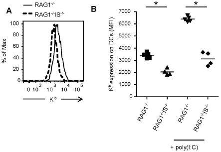
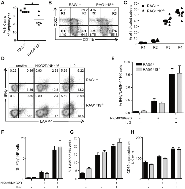
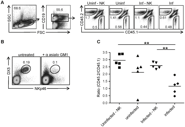
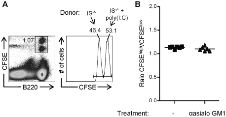
Natural killer (NK) cells are part of the innate immune system and contribute to the eradication of virus infected cells and tumors. NK cells express inhibitory and activating receptors and their decision to kill a target cell is based on the balance of signals received through these receptors. MHC class I molecules are recognized by inhibitory receptors, and their presence during NK cell education influences the responsiveness of peripheral NK cells. We here demonstrate that mice with reduced MHC class I cell surface expression, due to deficiency of immunoproteasomes, have responsive NK cells in the periphery, indicating that the lower MHC class I levels do not alter NK cell education. Following adoptive transfer into wild-type (wt) recipients, immunoproteasome-deficient splenocytes are tolerated in naive but rejected in virus-infected recipients, in an NK cell dependent fashion. These results indicate that the relatively low MHC class I levels are sufficient to protect these cells from rejection by wt NK cells, but that this tolerance is broken in infection, inducing an NK cell-dependent rejection of immunoproteasome-deficient cells.


Conflict of interest statement

Competing Interests: The authors have declared that no competing interests exist.

Figures

Figure 1
Figure 1. Effect of immunoproteasome-deficiency on constitutive expression and activation-induced upregulation of MHC class I molecules.
Splenocytes of untreated or poly(I:C) treated RAG1−/− and RAG1−/−β2i/MECL-1−/−β5i/LMP7−/− (RAG1−/−IS−/−) mice were analyzed for the expression of H2-Kb on DCs (CD11chiCD11bint). (A) Representative histogram of untreated mice showing H2-Kb expression on DCs. (B) H2-Kb mean fluorescent intensity (MFI) on DCs of individual mice. Data are representative of three independent experiments, n = 4–6 mice per group. Statistical analysis was performed using a Mann-Whitney U test. *, P<0.05.
Figure 2
Figure 2. Immunoproteasome-deficient mice have responsive peripheral NK cells.
(A–C) Splenocytes of RAG1−/− and RAG1−/−β2i/MECL-1−/−β5i/LMP7−/− (RAG1−/−IS−/−) mice were analyzed by flow cytometry (A) Frequencies of NK cells (DX5+NKp46+) as percentage of total lymphocytes. (B,C) Expression of CD11b and CD27 on DX5+NKp46+ NK cells. (B) Representative FACS plots and (C) graph showing percentages of subsets for individual mice for the regions indicated in (B). (D–H) Splenocytes of RAG1−/− and RAG1−/−IS−/− mice were incubated on plates coated with anti-NKG2D and anti-NKp46, in the presence or absence of IL-2, or left unstimulated. Frequencies of IFNγ+, LAMP-1+ NK cells and CD69 mean fluorescent intensity (MFI) on NK cells were determined by flow cytometry. (D) Representative FACS plots showing IFNγ+ and LAMP-1+ NK cells. (E–H) Bars showing the percentages of IFNγ+LAMP-1+ NK cells (E), IFNγ+ NK cells (F), LAMP-1+ NK cells (G), and CD69 MFIs on NK cells (H). Results are shown as mean ± S.E.M. Data are representative for 2 independent experiments with 6 mice per group. Statistical analysis was performed using Mann-Whitney U test. *, P<0.05.
Figure 3
Figure 3. Rejection of immunosubunit-deficient cells in influenza virus infected mice.
Splenocytes of congenic CD45.1 (wt) and CD45.2 β2i/MECL-1−/−β5i/LMP7−/− (IS−/−) mice were 1∶1 mixed based on total cell numbers, and transferred i.v. into untreated or anti-asialo GM1 treated CD45.1.2 recipients, that were subsequently infected i.n. with influenza virus or left uninfected. Recipient mice were sacrificed 8 days later and spleens were analyzed. (A) Representative FACS plots showing gating strategies and percentages of recovered CD45.1+ (wt) and CD45.2+ (IS−/−) cells of CD19+ cells in the different groups: uninf – NK (uninfected, anti-asialo GM1 treated), uninf (uninfected, untreated), inf – NK (influenza virus infected, anti-asialo GM1 treated), inf (influenza virus infected, untreated). (B) Representative FACS plots gated on TCRβ cells showing staining for DX5 and NKp46 on splenocytes from untreated and anti-asialo GM1 treated mice. Cells in the gate are DX5+NKp46+ NK cells. (C) Ratio of CD45.2 (IS−/−)/CD45.1 (wt) calculated by dividing absolute numbers of CD19+ CD45.2+ (IS−/−) cells by absolute numbers of CD19+ CD45.1+ (wt) cells. Results are representative for 2 independent experiments with 5–6 mice per group. Statistical analysis was performed using a Mann-Whitney U test. **, P<0.01.
Figure 4
Figure 4. Adoptively transferred splenocytes of poly(I:C)-treated immunoproteasome-deficient mice are tolerated by NK cells in naïve wt recipients.
CFSE-labeled splenocytes of untreated β2i/MECL-1−/−β5i/LMP7−/− (IS−/−; CFSElow) and of poly(I:C) treated IS−/− (CFSEhigh) mice were mixed 1∶1 and transferred i.v. into untreated or anti-asialo GM1 treated wt recipients. Recipient mice were sacrificed 17 h later and spleens were analyzed. (A) Representative FACS plots showing gating strategies. (B) Ratios of poly(I:C) treated (CFSEhigh) and untreated (CFSElow) IS−/− cells calculated by dividing absolute numbers of B220+CFSEhigh cells by absolute numbers of B220+CFSElow cells. Results represent one experiment with 6 mice per group.

Similar articles

Cited by

References

    1. Lanier LL. Up on the tightrope: Natural killer cell activation and inhibition. Nat Immunol. 2008;9(5):495–502. - PMC - PubMed
    1. Yokoyama WM, Altfeld M, Hsu KC. Natural killer cells: Tolerance to self and innate immunity to viral infection and malignancy. Biol Blood Marrow Transplant. 2010;16(1 Suppl):S97–S105. - PMC - PubMed
    1. Karre K, Ljunggren HG, Piontek G, Kiessling R. Selective rejection of H-2-deficient lymphoma variants suggests alternative immune defence strategy. Nature. 1986;319(6055):675–678. - PubMed
    1. Kim S, Poursine-Laurent J, Truscott SM, Lybarger L, Song YJ, et al. Licensing of natural killer cells by host major histocompatibility complex class I molecules. Nature. 2005;436(7051):709–713. - PubMed
    1. Fernandez NC, Treiner E, Vance RE, Jamieson AM, Lemieux S, et al. A subset of natural killer cells achieves self-tolerance without expressing inhibitory receptors specific for self-MHC molecules. Blood. 2005;105(11):4416–4423. - PMC - PubMed

Publication types

MeSH terms

Substances