

Identification and characterization of peripheral T-cell lymphoma-associated SEREX antigens
- PMID: 21887344
- PMCID: PMC3161784
- DOI: 10.1371/journal.pone.0023916
Identification and characterization of peripheral T-cell lymphoma-associated SEREX antigens
Abstract
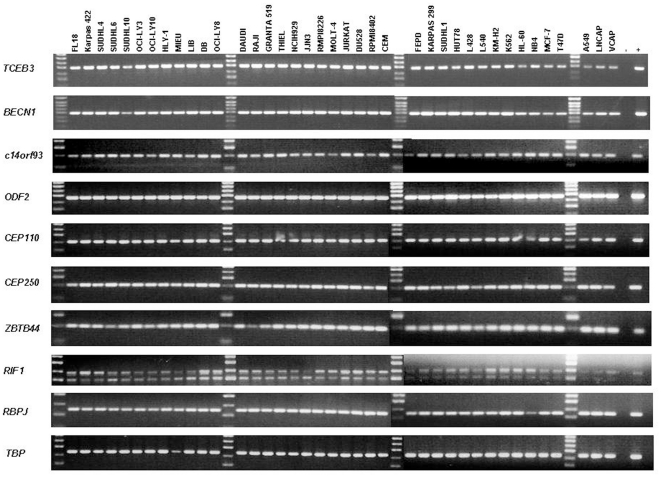
Peripheral T-cell lymphomas (PTCL) are generally less common and pursue a more aggressive clinical course than B-cell lymphomas, with the T-cell phenotype itself being a poor prognostic factor in adult non-Hodgkin lymphoma (NHL). With notable exceptions such as ALK(+) anaplastic large cell lymphoma (ALCL, ALK+), the molecular abnormalities in PTCL remain poorly characterised. We had previously identified circulating antibodies to ALK in patients with ALCL, ALK(+). Thus, as a strategy to identify potential antigens associated with the pathogenesis of PTCL, not otherwise specified (PTCL, NOS), we screened a testis cDNA library with sera from four PTCL, NOS patients using the SEREX (serological analysis of recombinant cDNA expression libraries) technique. We identified nine PTCL, NOS-associated antigens whose immunological reactivity was further investigated using sera from 52 B- and T-cell lymphoma patients and 17 normal controls. The centrosomal protein CEP250 was specifically recognised by patients sera and showed increased protein expression in cell lines derived from T-cell versus B-cell malignancies. TCEB3, BECN1, and two previously uncharacterised proteins, c14orf93 and ZBTB44, were preferentially recognised by patients' sera. Transcripts for all nine genes were identified in 39 cancer cell lines and the five genes encoding preferentially lymphoma-recognised antigens were widely expressed in normal tissues and mononuclear cell subsets. In summary, this study identifies novel molecules that are immunologically recognised in vivo by patients with PTCL, NOS. Future studies are needed to determine whether these tumor antigens play a role in the pathogenesis of PTCL.
Conflict of interest statement
Figures


References
-
- The Non-Hodgkin's Lymphoma Classification Project. A clinical evaluation of the International Lymphoma Study Group classification of non-Hodgkin's lymphoma. Blood. 1997;89:3909–3918. - PubMed
-
- Swerdlow SH, Campo E, Harris NL, Jaffe ES, Pileri SA, et al., editors. WHO Classification of Tumours of Haematopoietic and Lymphoid Tissues. Lyon: IARC; 2008.
-
- Jaffe E, Harris N, Stein H, Vardiman J. Pathology and genetics: tumours of haematopoietic and lymphoid tissues. In: Kleihues P, Sobin L, editors. World Health Organization Classification of Tumours. Lyon: IARC Press; 2001. pp. 190–235.
-
- Went P, Agostinelli C, Gallamini A, Piccaluga PP, Ascani S, et al. Marker expression in peripheral T-cell lymphoma: a proposed clinical-pathologic prognostic score. J Clin Oncol. 2006;24:2472–2479. - PubMed
-
- Lepretre S, Buchonnet G, Stamatoullas A, Lenain P, Duval C, et al. Chromosome abnormalities in peripheral T-cell lymphoma. Cancer Genet Cytogenet. 2000;117:71–79. - PubMed
Publication types
MeSH terms
Substances
LinkOut - more resources
Full Text Sources
