

IL-17A expression is localised to both mononuclear and polymorphonuclear synovial cell infiltrates
- PMID: 21887369
- PMCID: PMC3161104
- DOI: 10.1371/journal.pone.0024048
IL-17A expression is localised to both mononuclear and polymorphonuclear synovial cell infiltrates
Abstract
Introduction: This study examines the expression of IL-17A-secreting cells within the inflamed synovium and the relationship to in vivo joint hypoxia measurements.
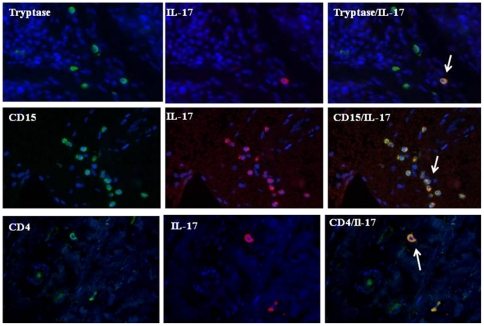
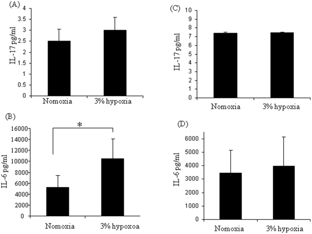
Methods: IL-17A expression was quantified in synovial tissue (ST), serum and synovial fluid (SF) by immunohistochemistry and MSD-plex assays. IL-6 SF and serum levels were measured by MSD-plex assays. Dual immunofluorescence for IL-17A was quantified in ST CD15+ cells (neutrophils), Tryptase+ (mast cells) and CD4+ (T cells). Synovial tissue oxygen (tpO(2)) levels were measured under direct visualisation at arthroscopy. Synovial infiltration was assessed using immunohistochemistry for cell specific markers. Peripheral blood mononuclear and polymorphonuclear cells were isolated and exposed to normoxic or 3% hypoxic conditions. IL-17A and IL-6 were quantified as above in culture supernatants.
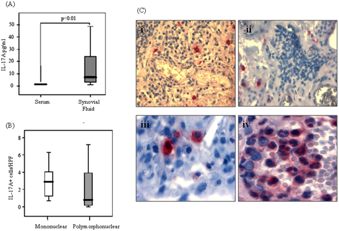
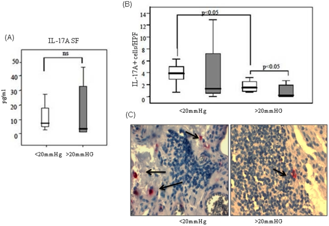
Results: IL-17A expression was localised to mononuclear and polymorphonuclear (PMN) cells in inflamed ST. Dual immunoflourescent staining co-localised IL-17A expression with CD15+ neutrophils Tryptase+ mast cells and CD4+T cells. % IL-17A positivity was highest on CD15+ neutrophils, followed by mast cells and then CD4+T-cells. The number of IL-17A-secreting PMN cells significantly correlated with sublining CD68 expression (r = 0.618, p<0.01). IL-17A SF levels correlated with IL-6 SF levels (r = 0.675, p<0.01). Patients categorized according to tp0(2)< or >20 mmHg, showed those with low tp0(2)<20 mmHg had significantly higher IL-17A+ mononuclear cells with no difference observed for PMNs. Exposure of mononuclear and polymorphonuclear cells to 3% hypoxia, significantly induced IL-6 in mononuclear cells, but had no effect on IL-17A expression in mononuclear and polymorphonuclear cells.
Conclusion: This study demonstrates IL-17A expression is localised to several immune cell subtypes within the inflamed synovial tissue, further supporting the concept that IL-17A is a key mediator in inflammatory arthritis. The association of hypoxia with Il-17A expression appears to be indirect, probably through hypoxia-induced pro-inflammatory pathways and leukocyte influx within the joint microenvironment.
Conflict of interest statement
Figures
Similar articles
-
Heterogeneous expression pattern of interleukin 17A (IL-17A), IL-17F and their receptors in synovium of rheumatoid arthritis, psoriatic arthritis and osteoarthritis: possible explanation for nonresponse to anti-IL-17 therapy?Arthritis Res Ther. 2014 Aug 22;16(4):426. doi: 10.1186/s13075-014-0426-z. Arthritis Res Ther. 2014. PMID: 25146432 Free PMC article.
-
IL-27-producing CD14(+) cells infiltrate inflamed joints of rheumatoid arthritis and regulate inflammation and chemotactic migration.Cytokine. 2011 Aug;55(2):237-44. doi: 10.1016/j.cyto.2011.04.020. Epub 2011 May 17. Cytokine. 2011. PMID: 21592822
-
Low dose methotrexate decreases intraarticular prostaglandin and interleukin 1 levels in antigen induced arthritis in rabbits.J Rheumatol. 1996 Dec;23(12):2092-7. J Rheumatol. 1996. PMID: 8970046
-
Synovial tissue hypoxia and inflammation in vivo.Ann Rheum Dis. 2010 Jul;69(7):1389-95. doi: 10.1136/ard.2009.119776. Epub 2010 May 3. Ann Rheum Dis. 2010. PMID: 20439288 Free PMC article.
-
Inside the Joint of Inflammatory Arthritis Patients: Handling and Processing of Synovial Tissue Biopsies for High Throughput Analysis.Front Med (Lausanne). 2022 Mar 14;9:830998. doi: 10.3389/fmed.2022.830998. eCollection 2022. Front Med (Lausanne). 2022. PMID: 35372383 Free PMC article. Review.
Cited by
-
[Hypoxia is a key factor in the inflammatory milieu of rheumatic diseases].Z Rheumatol. 2012 Jan;71(1):64-7. doi: 10.1007/s00393-011-0782-x. Z Rheumatol. 2012. PMID: 22143445 German.
-
Hypoxia, mitochondrial dysfunction and synovial invasiveness in rheumatoid arthritis.Nat Rev Rheumatol. 2016 Jul;12(7):385-97. doi: 10.1038/nrrheum.2016.69. Epub 2016 May 26. Nat Rev Rheumatol. 2016. PMID: 27225300 Review.
-
Neutrophils with low production of reactive oxygen species are activated during immune priming and promote development of arthritis.Redox Biol. 2024 Dec;78:103401. doi: 10.1016/j.redox.2024.103401. Epub 2024 Oct 18. Redox Biol. 2024. PMID: 39471640 Free PMC article.
-
The correlations between IL-17 vs. Th17 cells and cancer patient survival: a systematic review.Oncoimmunology. 2015 Mar 6;4(2):e984547. doi: 10.4161/2162402X.2014.984547. eCollection 2015 Feb. Oncoimmunology. 2015. PMID: 25949881 Free PMC article. Review.
-
Cyr61 is involved in neutrophil infiltration in joints by inducing IL-8 production by fibroblast-like synoviocytes in rheumatoid arthritis.Arthritis Res Ther. 2013;15(6):R187. doi: 10.1186/ar4377. Arthritis Res Ther. 2013. PMID: 24517278 Free PMC article.
References
-
- Combe B. Progression in early rheumatoid arthritis. Best Pract Res Clin Rheumatol. 2009;23:59–69. - PubMed
-
- Veale DJ, Maple C. Cell adhesion molecules in rheumatoid arthritis. Drugs Aging. 1996;9:87–92. - PubMed
-
- Iwamoto T, Okamoto H, Toyama Y, Momohara S. Molecular aspects of rheumatoid arthritis: chemokines in the joints of patients. FEBS Journal. 2008;275:4448–4455. - PubMed
Publication types
MeSH terms
Substances
LinkOut - more resources
Full Text Sources
Research Materials
