. 2011;6(8):e24154.
doi: 10.1371/journal.pone.0024154. Epub 2011 Aug 26.

RssAB signaling coordinates early development of surface multicellularity in Serratia marcescens

Affiliations

RssAB signaling coordinates early development of surface multicellularity in Serratia marcescens

Yu-Huan Tsai et al. PLoS One. 2011.

Abstract

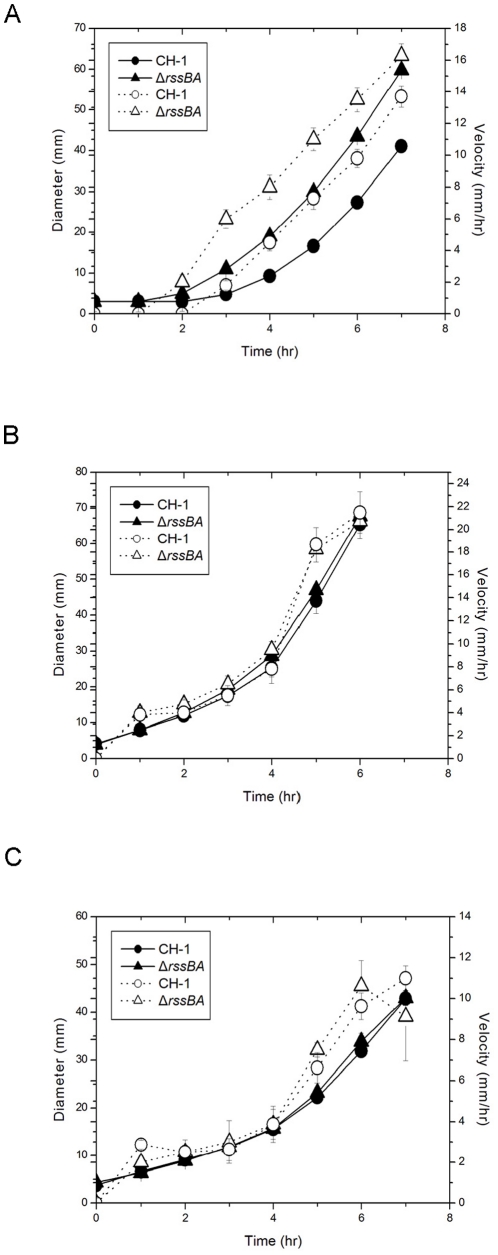
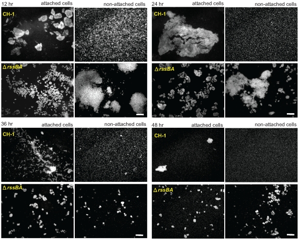
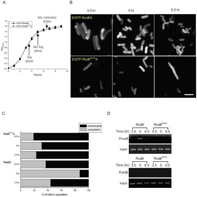
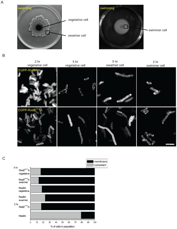
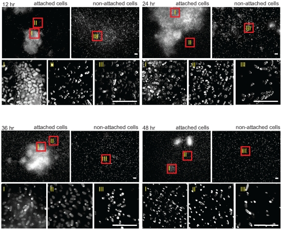
Bacteria can coordinate several multicellular behaviors in response to environmental changes. Among these, swarming and biofilm formation have attracted significant attention for their correlation with bacterial pathogenicity. However, little is known about when and where the signaling occurs to trigger either swarming or biofilm formation. We have previously identified an RssAB two-component system involved in the regulation of swarming motility and biofilm formation in Serratia marcescens. Here we monitored the RssAB signaling status within single cells by tracing the location of the translational fusion protein EGFP-RssB following development of swarming or biofilm formation. RssAB signaling is specifically activated before surface migration in swarming development and during the early stage of biofilm formation. The activation results in the release of RssB from its cognate inner membrane sensor kinase, RssA, to the cytoplasm where the downstream gene promoters are located. Such dynamic localization of RssB requires phosphorylation of this regulator. By revealing the temporal activation of RssAB signaling following development of surface multicellular behavior, our findings contribute to an improved understanding of how bacteria coordinate their lifestyle on a surface.


Conflict of interest statement

Competing Interests: The authors have declared that no competing interests exist.

Figures

Figure 1
Figure 1. RssAB TCS regulates duration of swarming lag period.
Swarming (A) and swimming (C) motility assay of S. marcescens CH-1 and isogenic mutant ΔrssBA were performed. (B) Swarmer cells harvested from the swarming edge when swarming diameter reached 50 mm were re-inoculated onto another fresh swarming plate at the original density. Diameters (solid line with closed symbols) and velocity (dotted line with open symbols) were shown. Overnight bacterial LB broth culture (1 µl, 1010 CFU/ml) was inoculated onto or into the centers of 0.8% (wt/vol) and 0.3% (wt/vol) Eiken agar LB plates at 30°C for swarming and swimming assay, respectively. Results shown were averages of measurements from four independent experiments.
Figure 2
Figure 2. RssAB regulates biofilm structure.
Bacteria were cultured in LB medium supplemented with 1% (wt/vol) sucrose and 0.05% (wt/vol) arabinose to overproduce EGFP for observation. Petri dishes with glass coverslips and bacterial suspension were incubated under 50 rpm shaking at 30°C. Biofilm cells attached on glass coverslips or non-attached cells in the broth were observed under fluorescence microscopy at time points indicated. No obvious cell clustering was observed at 6 hr in both attached cells and non-attached cells of S. marcescens CH-1 and ΔrssBA (data not shown). Scale bar, 50 µm.
Figure 3
Figure 3. In vitro interaction between RssB and cRssA.
GST, GST-RssB and GST-RssBD51E were pretreated with Ac-P, followed by incubation with His-cRssA and glutathione sepharose-4B in interaction buffer. After four times wash with interaction buffer, SDS-PAGE sample buffer was added to each reaction and separated by 12% SDS-PAGE. Proteins were detected by coomassie brilliant blue stain (upper) or anti-hexa-Histidine monoclonal antibody (lower). The molecular weights of GST-RssB and His-cRssA are approximately 50 kDa and 30 kDa respectively. The relative amount of His-cRssA being pulled-down compared to the input one was indicated.
Figure 4
Figure 4. Directly imaging the localization of EGFP-RssB in LB broth culture.
(A) The growth dynamic of S. marcescens CH-1 harboring pEGFP-RssBA(Sm) or pEGFP-RssBD51EA(Sm) was shown. (B) EGFP-RssB was localized at cell membrane in the log (3.5 hr) and the late stationary (6.5 hr) growth phases, and became dispersed in the cytoplasm in the late log phase (5hr) in LB broth culture when comparable amount of RssA was co-expressed. Scale bar, 2 µm (C) The percentage of cells showing EGFP-RssB localizing at the cell membrane or in the cytoplasm. At least 200 cells were counted for each assay condition. For EGFP-RssBA, both EGFP-RssB and RssA were co-expressed under PBAD promoter control and induced by 0.1% (wt/vol) arabinose. EGFP-RssBD51EA production was achieved in the same manner. (D) Binding of RssB∼P to its own promoter upon dispersing to the cytoplasm was demonstrated. In vivo target DNA binding properties of RssB and RssBD51E were examined by mChIP assay. Captured and total (input) DNA were subjected to PCR using primers PrssBFootF/PrssBFootR and BPshlBF/BPshlBR specific to amplify PrssB and PshlB, respectively. As RssB∼P cannot directly bind PshlB in vitro , PshlB was used as a negative control.
Figure 5
Figure 5. Cytolocalization of EGFP-RssB during swarming development.
(A) The patterns of swarming and swimming after 5 hr of inoculation were shown. To image the cytolocalization of EGFP fusion protein in different cell populaiton, cells were taken from the zones of interest as indicated. For the vegetative and swarmer cells, cells were taken from the surface of plates, whereas the swimmer cells were taken from within the agar per se. (B) EGFP-RssB was dispersed in the cytoplasm in central, non-motile cells before initiation of surface migration, and homogenously localized at the periphery of the cell after migration (5 hr vegetative cell and 5 hr swarmer cell) following swarming development. The localization of EGFP-RssB at the cell membrane was also observed in the cells during swimming (2 hr swimmer cell). (C) The percentage of two types of EGFP-RssB localization pattern in cell population cultured on swarming plates was shown. At least 200 cells were counted for each assay condition. For EGFP-RssBA, both EGFP-RssB and RssA were co-expressed under PBAD promoter control and induced by 0.1% (wt/vol) arabinose. EGFP-RssBD51EA production was achieved in the same manner. Swarming was performed at 30°C and the bacteria were directly picked by tip and immobilized between a coverslip and a thin slab of 1.5% (wt/vol) agarose for microscopy. Scale bar, 2 µm.
Figure 6
Figure 6. Cytolocalization of EGFP-RssB during biofilm development.
Samples were prepared as described in Fig. 2 with cells harboring pEGFP-RssBA(Sm) and observed under 100X objective lens. RssAB signaling status of the cells aggregated on the surface (I), attached on the surface individually (II) and non-attached cells (III) were shown. EGFP-RssB is localized at the cell membrane in both attached cells and non-attached cells following biofilm development except for the 12 hr attached cells in which EGFP-RssB evenly distributed in the cytoplasm when aggregating on the surface. Scale bar, 5 µm.

References

    1. Eberl L, Molin S, Givskov M. Surface motility of Serratia liquefaciens MG1. J Bacteriol. 1999;181:1703–1712. - PMC - PubMed
    1. Rather PN. Swarmer cell differentiation in Proteus mirabilis. Environ Microbiol. 2005;7:1065–1073. - PubMed
    1. Stoodley P, Sauer K, Davies DG, Costerton JW. Biofilms as complex differentiated communities. Annu Rev Microbiol. 2002;56:187–209. - PubMed
    1. Verstraeten N, Braeken K, Debkumari B, Fauvart M, Fransaer J, et al. Living on a surface: swarming and biofilm formation. Trends Microbiol. 2008;16:496–506. - PubMed
    1. Butler MT, Wang Q, Harshey RM. Cell density and mobility protect swarming bacteria against antibiotics. Proc Natl Acad Sci U S A. 2010;107:3776–3781. - PMC - PubMed

Publication types

Substances