. 2011 Oct 7;10(10):4547-55.
doi: 10.1021/pr200355w. Epub 2011 Sep 22.

Myosin binding protein-C slow is a novel substrate for protein kinase A (PKA) and C (PKC) in skeletal muscle

Affiliations

Myosin binding protein-C slow is a novel substrate for protein kinase A (PKA) and C (PKC) in skeletal muscle

Maegen A Ackermann et al. J Proteome Res. .

Abstract

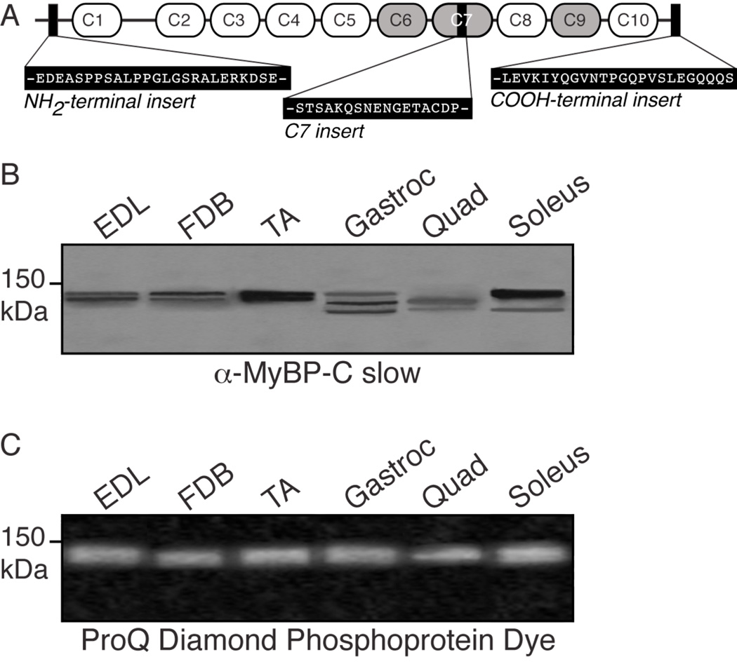
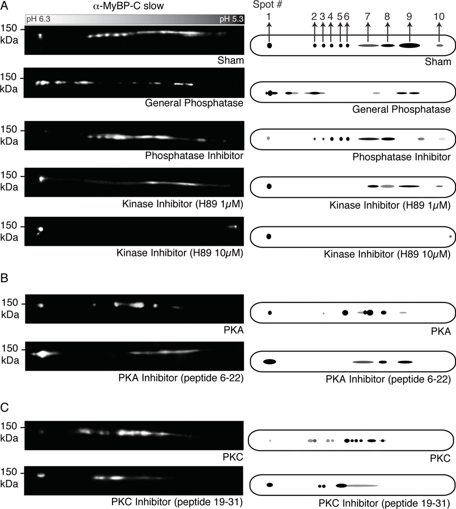
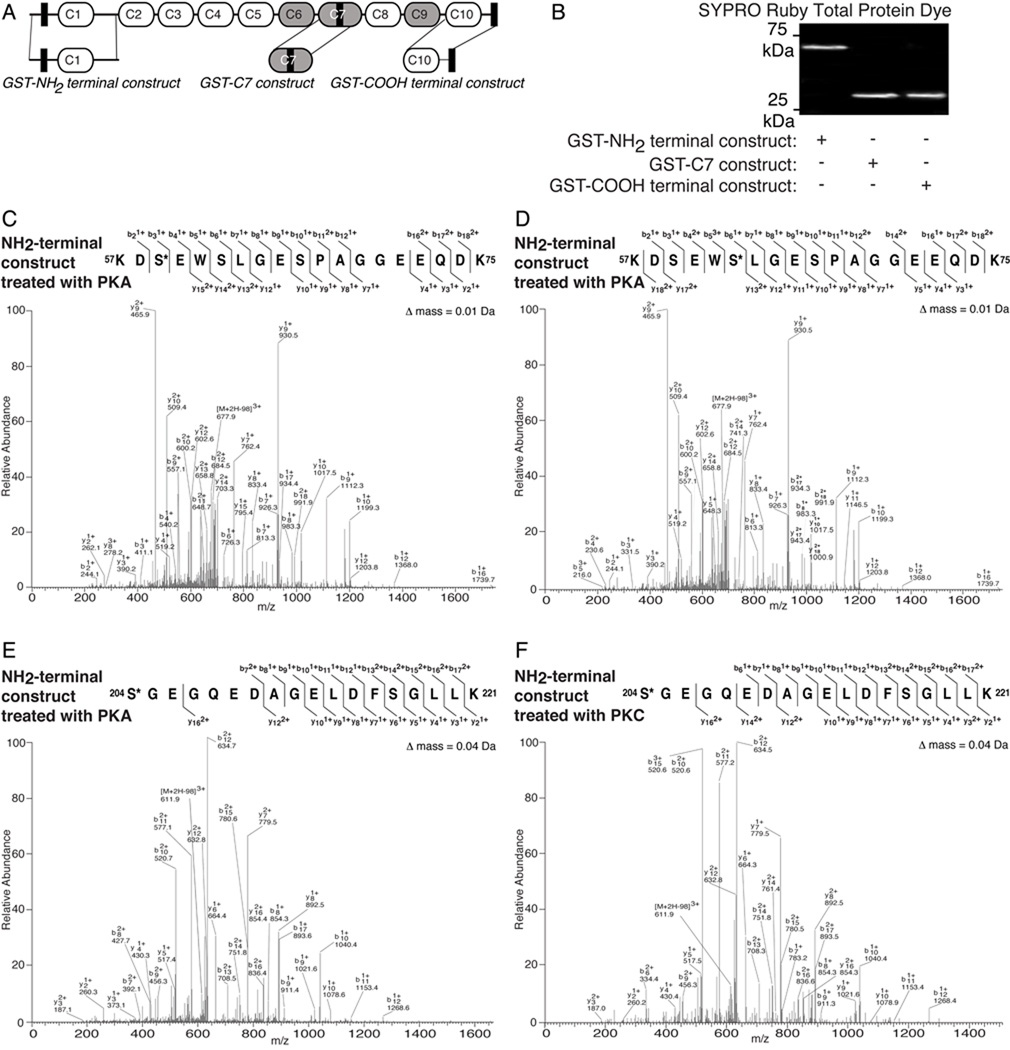
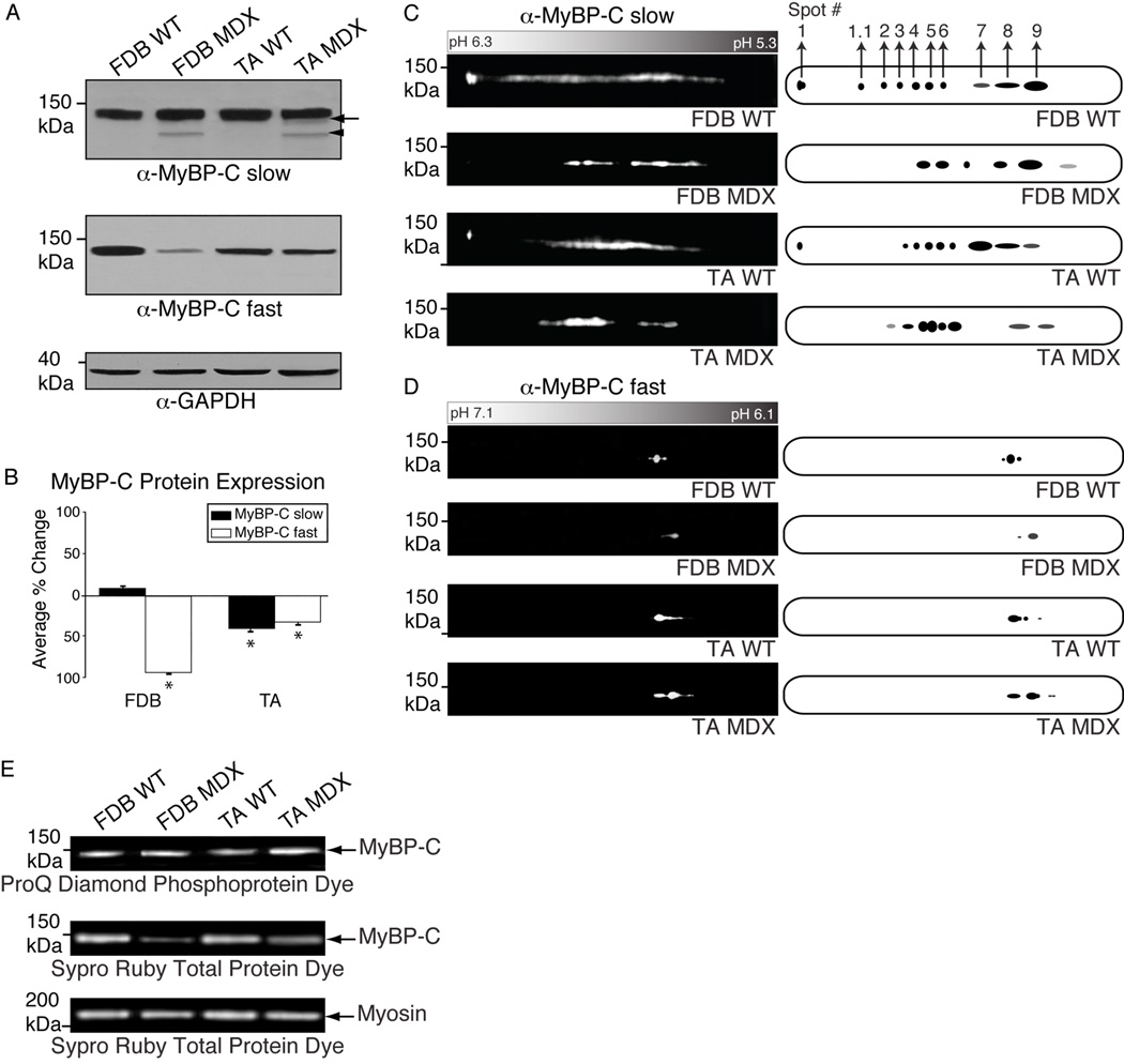
Myosin Binding Protein-C slow (MyBP-C slow), a family of thick filament-associated proteins, consists of four alternatively spliced forms, namely variants 1-4. Variants 1-4 share common structures and sequences; however, they differ in three regions: variants 1 and 2 contain a novel 25-residue long insertion at the extreme NH(2)-terminus, variant 3 carries an 18-amino acid long segment within immunoglobulin (Ig) domain C7, and variant 1 contains a unique COOH-terminus consisting of 26-amino acids, while variant 4 does not possess any of these insertions. Variants 1-4 are expressed in variable amounts among skeletal muscles, exhibiting different topographies and potentially distinct functions. To date, the regulatory mechanisms that modulate the activities of MyBP-C slow are unknown. Using an array of proteomic approaches, we show that MyBP-C slow comprises a family of phosphoproteins. Ser-59 and Ser-62 are substrates for PKA, while Ser-83 and Thr-84 are substrates for PKC. Moreover, Ser-204 is a substrate for both PKA and PKC. Importantly, the levels of phosphorylated skeletal MyBP-C proteins (i.e., slow and fast) are notably increased in mouse dystrophic muscles, even though their overall amounts are significantly decreased. In brief, our studies are the first to show that the MyBP-C slow subfamily undergoes phosphorylation, which may regulate its activities in normalcy and disease.


Figures

Figure 1
Figure 1
Adult murine skeletal muscles express multiple MyBP-C slow variants. A: Schematic representation of a “hypothetical” murine MyBP-C slow transcript consisting of tandem Ig (white ovals) and FnIII (grey ovals) domains with the three novel insertions shown (black rectangles). B–C: Protein homogenates prepared from murine skeletal muscles were labeled with anti-MyBP-C slow antibody (B) and stained with ProQ Diamond Dye that specifically detects phosphorylated proteins (C). At least three immunoreactive bands were detected, ranging in size from ~125–135 kDa that correspond to the different variants of MyBP-C slow (v1–v4; please see text). Notably, a broad band of ~125–135 kDa was detected by the ProQ Diamond phospho-dye in all skeletal muscles tested.
Figure 2
Figure 2
Proteomic analysis of MyBP-C slow in fast twitch FDB muscle. A–C: Protein homogenates prepared from murine FDB skeletal muscle were subjected to different phospho-chemical treatments, analyzed by 2-D gel electrophoresis and immunostained with a MyBP-C slow antibody (A). Representative blots are shown on the left with corresponding cartoon representations on the right. MyBP-C slow appears to be a substrate of PKA (B) and PKC (C) in vivo.
Figure 3
Figure 3
MyBP-C slow is phosphorylated by PKA and PKC in vitro. A: Schematic representation of the same “hypothetical” protein shown in Fig. 1A, highlighting recombinant MyBP-C slow constructs. Recombinant polypeptides are denoted as GST-NH2 terminal construct, GST-C7 construct and GST-COOH terminal construct and detected by SYPRO ruby total protein stain (B). C–F: Phosphorylation events mediated by PKA and PKC were identified in the GST-NH2-terminal construct via mass spectrometry analysis using LTQ-orbitrap. MS/MS scans of peptides carrying phosphorylated Ser-59 by PKA (C), Ser-62 by PKA (D) and Ser-204 by PKA (E) and PKC (F), identified by Bioworks-SEQUEST, with Xcorr scores of 4.471, 4.757, 4.142 and 4.475, respectively, and manually validated are shown.
Figure 4
Figure 4
Examination of the expression profile of MyBP-C slow and fast in dystrophic (MDX) skeletal muscles. A: Protein homogenates prepared from FDB and TA skeletal muscles from wild type (WT) and MDX mice were separated by 1-D SDS-PAGE and probed with antibodies to MyBP-C slow (A, top panel) and fast (A, middle panel). An immunoreactive band of ~125 kDa representing MyBP-C slow v4 (arrowhead) was detected in dystrophic FDB and TA, but not wild type, tissues. Moreover, the ~128–129 kDa band, corresponding to v2 and/or v3, is significantly diminished in MDX TA muscle (arrow). Similarly, the amounts of MyBP-C fast are considerably reduced in dystrophic FDB and TA muscles. Equal loading of protein lysates was confirmed by immunostaining for GAPDH (A, bottom panel). B: Densitometric analysis of immunoreactive bands was used to estimate the relative amounts of MyBP-C slow (black bars) and fast (white bars) in wild type and dystrophic muscles; differences in expression levels are reported as percent changes (p<0.01; Students t-test). C–D: The same homogenates were analyzed by 2-D SDS-PAGE and immunostained for MyBP-C slow (C) and fast (D). A distinct shift of both MyBP-C slow and fast immunoreactive spots toward a more phosphorylated state was observed in dystrophic muscles. E: Consistent with this, the ~125–135 kDa band, encompassing skeletal MyBP-C slow and fast, was of higher intensity in homogenates prepared from dystrophic FDB and TA muscles compared to the band detected in the respective normal muscles after staining with ProQ Diamond phosphospecific dye (E, top panel), albeit the total amounts of skeletal MyBP-C protein were significantly reduced, as indicated by Sypro Ruby staining (E, middle panel); myosin staining with Sypro Ruby was used as a loading control (E, bottom panel), indicating equivalent levels between wild type and dystrophic muscles.

References

    1. Oakley CE, Chamoun J, Brown LJ, Hambly BD. Myosin binding protein-C: enigmatic regulator of cardiac contraction. Int J Biochem Cell Biol. 2007;39(12):2161–2166. - PubMed
    1. Gautel M, Castiglione Morelli MA, Pfuhl M, Motta A, Pastore A. A calmodulin-binding sequence in the C-terminus of human cardiac titin kinase. Eur J Biochem. 1995;230(2):752–759. - PubMed
    1. Weber FE, Vaughan KT, Reinach FC, Fischman DA. Complete sequence of human fast-type and slow-type muscle myosin-binding-protein C (MyBP-C). Differential expression, conserved domain structure and chromosome assignment. Eur J Biochem. 1993;216(2):661–669. - PubMed
    1. Kang LH, Rughani A, Walker ML, Bestak R, Hoh JF. Expression of masticatory-specific isoforms of myosin heavy-chain, myosin-binding protein-C and tropomyosin in muscle fibers and satellite cell cultures of cat masticatory muscle. J Histochem Cytochem. 2010;58(7):623–634. - PMC - PubMed
    1. Kang LH, Hoh JF. Regulation of jaw-specific isoforms of myosin-binding protein-C and tropomyosin in regenerating cat temporalis muscle innervated by limb fast and slow motor nerves. J Histochem Cytochem. 2010;58(11):989–1004. - PMC - PubMed

Publication types