. 2011 Sep 5:11:15.
doi: 10.1186/1471-2253-11-15.

Esmolol modulates inhibitory neurotransmission in the substantia gelatinosa of the spinal trigeminal nucleus of the rat

Affiliations

Esmolol modulates inhibitory neurotransmission in the substantia gelatinosa of the spinal trigeminal nucleus of the rat

Yutaka Yasui et al. BMC Anesthesiol. .

Abstract

Background: β1-adrenaline receptor antagonists are often used to avoid circulatory complications during anesthesia in patients with cardiovascular diseases. Of these drugs, esmolol, a short-acting β antagonist, is also reported to exert antinociceptive and anesthetic sparing effects. This study was designed to identify the central mechanism underlying the antinociceptive effect of esmolol.

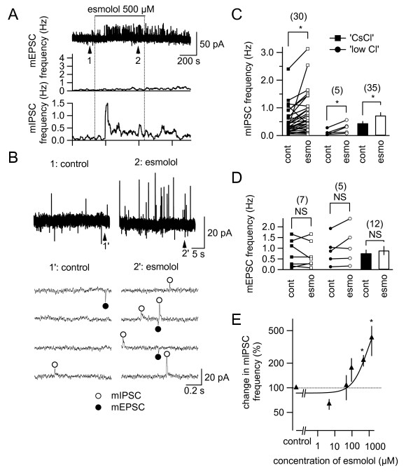
Methods: Wistar rats (7-21 d, 17-50 g) were anesthetized with ketamine (100-150 mg/kg) or isoflurane (5%) and decapitated. Horizontal slices (400-μm thick) of the lower brainstem containing the substantia gelatinosa (SG) of the caudal part of the spinal trigeminal nucleus (Sp5c), in which the nociceptive primary afferents form the first intracranial synapses, were made with a vibrating slicer. The miniature inhibitory and excitatory postsynaptic currents (mIPSCs and mEPSCs, respectively) were simultaneously recorded from visually identified SG neurons of the Sp5c in the presence of tetrodotoxin (1 μM). Additionally, mIPSCs were recorded during pharmacological isolation of GABA- and glycine-mediated mIPSCs with kynurenic acid (1 mM).

Results: Esmolol (500 μM) significantly and selectively increased the mIPSC frequency (to 214.2% ± 34.2% of the control, mean ± SEM, n = 35; P < 0.001), but not that of mEPSCs, without changing their amplitude. The increase in mIPSC frequency with esmolol was not affected by prior activation of β receptors with isoproterenol (100 μM) but it was significantly attenuated by removal of extracellular Ca2+.

Conclusions: These data suggest that esmolol modulates inhibitory transmitter release in the Sp5c through a mechanism involving Ca2+-entry but in a β1-adrenoceptor-independent manner. The present results suggest that the facilitation of inhibitory transmitter release in the central nociceptive network underlies, at least in part, the antinociceptive effect of esmolol.


Figures

Figure 1
Figure 1
Selective increase in miniature inhibitory postsynaptic current (mIPSC) frequency by esmolol in neurons of the caudal part of the spinal trigeminal nucleus (Sp5c). (A) Top: the membrane current recording of an Sp5c neuron at a holding potential of -40 mV. Esmolol was applied at the horizontal bar. Middle: the time course of the changes in miniature excitatory postsynaptic current (mEPSC) frequency with esmolol. Bottom: the time course of the changes in mIPSC frequency with esmolol. The abscissae of these graphs are identical (time; 5 min/div). (B) 1 and 2 are time-extended traces taken at the points 1 and 2 in the trace in A (top). mIPSCs and mEPSCs (outward and inward events, respectively) are marked with open circles above the traces and filled circles below the traces, respectively. (C) and (D) Summaries of the effects of esmolol on mIPSC and mEPSC frequency, respectively. "cont", mPSC frequency observed in 3-min control period before esmolol application (filled markers and bars); "esmo", those at 10-min application of esmolol (500 μM; open markers and bars). The numbers in parentheses indicate the number of neurons tested and were used for the statistics. Left, results of recordings with "CsCl-based" internal solution (squares) under pharmacological isolation of mIPSCs (C) and mEPSCs (D); center, results of recordings with "low-Cl" internal solution in five cells (circles) in which mIPSCs (C) and mEPSCs (D) were simultaneously recorded (see Methods); right bars, pooled summaries based on the results with "CsCl" internal solution and "low-Cl" solution. *, P < 0.05; NS, not significantly different from pre-administration values (100%). Paired t-test. (E) Concentration-response relationship between esmolol and the changes in mIPSC frequency. The curve indicates the best-fit Hill equations for the data for esmolol. The number of neurons used to estimate the mean values and curve-fitting was 39 (control); 5 (5 μM); 5 (50 μM); 4 (100 μM); 35 (500 μM); 7 (1500 μM). *, P < 0.02; Mann-Whitney's U test; vs. control (no drug application). The horizontal broken line indicates the control values (100%).
Figure 2
Figure 2
Effects of β receptor agonists on the effect of esmolol and the effects of another beta antagonist, landiolol, on synaptic inhibitory transmission in the caudal part of the spinal trigeminal nucleus (Sp5c) neurons. (A and B) Top: the membrane current recording of a neuron of the Sp5c in the absence (A) and presence (B) of 10-min prior administration of isoproterenol (100 μM). Recordings with "CsCl-based" internal solution. Esmolol was applied at the horizontal bar. Bottom of A and B: time-expanded continuous traces taken at points 1 and 2 in the top trace. (C) Summary of the effect of esmolol on mIPSC frequency in the absence (open circles; filled bar) and presence (filled circles; open bar) of isoproterenol. The bars show the average values. *, P < 0.05; Mann Whitney's U-test. NS, not significantly different; vs. pre-esmolol values. Each circle represents the data from one neuron (n = 6 neurons). (D) Top: the membrane current recording of a neuron of Sp5c using "CsCl-based" internal solution. Landiolol was applied at the horizontal bar. Bottom: time-expanded continuous traces taken at points 1 and 2 in the top trace. (E) Concentration-response relationship between landiolol and the changes in mIPSC frequency. The number of neurons used to estimate the mean values was 12 (control); 4 (5 μM); 4 (50 μM); 4 (100 μM); 10 (500 μM); 4 (1500 μM). Mann-Whitney's U test; vs. control (no drug application). The horizontal broken line indicates the control values (100%).
Figure 3
Figure 3
The increase in miniature inhibitory postsynaptic current (mIPSC) frequency by esmolol was dependent on extracellular Ca2+. (A and B) Top: the membrane current recording of a neuron of the caudal part of the spinal trigeminal nucleus in the presence (A) and in the absence (B) of extracellular Ca2+. Esmolol (500 μM) was applied at the horizontal bars. Recordings with "CsCl-based" internal solution. Bottom of A and B: time-expanded continuous traces taken at points 1 and 2 indicated in the top traces showing the control and the peak effects of drugs. (C) Summery of the effects of esmolol on mIPSC frequency. *, P < 0.001; Mann Whitney's U-test. NS, not significantly different; vs. pre-administration control values. The numbers in the bars indicate the number of neurons analyzed. The horizontal broken lines indicate the control values (100%).
Figure 4
Figure 4
The increase in spontaneous inhibitory postsynaptic current (sIPSC) frequency by esmolol in neurons of the caudal part of the spinal trigeminal nucleus (Sp5c). (A) The membrane current recording of an Sp5c neuron. Esmolol was applied at the horizontal bar. (B) Traces showing evoked IPSC (eIPSC) waveforms (average of eight consecutive traces) evoked by stimulation before (1; left), during (2; middle) and after (3; right) esmolol application in the Sp5c. 1-3 were sampled at points 1-3 in A. (C) 1-3 are time-extended traces taken at points 1-3 in the trace in A. (D) and (E) Summary of the effects of esmolol on eIPSC amplitude and sIPSC frequency, respectively. *, P < 0.05; Mann-Whitney's U-test. (F) The time course of the changes with esmolol in sIPSC frequency (thick line) and eIPSC amplitude (thin line) that were simultaneously recorded. The abscissae of these graphs are identical (time).

References

    1. Davidson EM, Doursout MF, Szmuk P, Chelly JE. Antinociceptive and cardiovascular properties of esmolol following formalin injection in rats. Can J Anaesth. 2001;48:59–64. doi: 10.1007/BF03019816. - DOI - PubMed
    1. Johansen JW, Flaishon R, Sebel PS. Esmolol reduces anesthetic requirement for skin incision during propofol/nitrous oxide/morphine anesthesia. Anesthesiology. 1997;86:364–71. doi: 10.1097/00000542-199702000-00011. - DOI - PubMed
    1. Johansen JW, Schneider G, Windsor AM, Sebel PS. Esmolol potentiates reduction of minimum alveolar isoflurane concentration by alfentanil. Anesth Analg. 1998;87:671–6. - PubMed
    1. Chia YY, Chan MH, Ko NH, Liu K. Role of beta-blockade in anaesthesia and postoperative pain management after hysterectomy. Br J Anaesth. 2004;93:799–805. doi: 10.1093/bja/aeh268. - DOI - PubMed
    1. Kadoi Y, Saito S. Possible indications of beta-blockers in the perioperative period other than prevention of cardiac ischemia. J Anesth. 2010;24:81–95. doi: 10.1007/s00540-009-0865-x. - DOI - PubMed

LinkOut - more resources