Review
. 2011 Oct;21(5):590-6.
doi: 10.1016/j.sbi.2011.08.007. Epub 2011 Aug 31.

Post-translational regulation of signaling mucins

Affiliations
Review

Post-translational regulation of signaling mucins

Paul J Cullen. Curr Opin Struct Biol. 2011 Oct.

Abstract

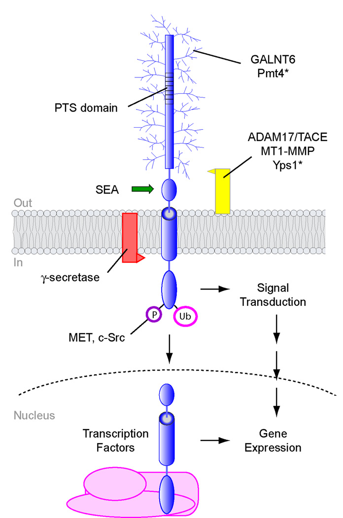
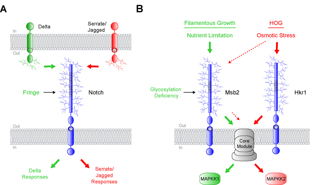
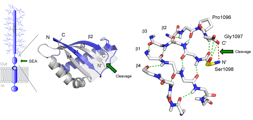
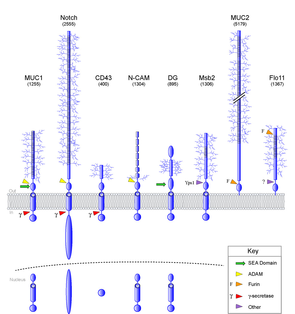
Signaling mucins are large transmembrane glycoproteins that regulate signal transduction pathways. Recent advances have shown that two major types of post-translational modifications, protein glycosylation and proteolytic processing, play important and unexpected roles in regulating signaling mucin function. New O-glycosyltransferases and proteases have been identified, and the structure of the domain that undergoes auto-proteolysis has been solved. A picture is beginning to emerge where specific glycosyl modifications and regulated processing control the signaling and adherence properties of signaling glycoproteins and contribute to the routing of signals to specific pathways.


Figures

Figure 1
Figure 1. Post-translational modifications of signaling mucins
A schematic representation of a signaling mucin is shown. Proteins that modify signaling mucins include O-glycosyltransferases, like GALNT6 and Pmt4, and proteases γ-secretase, Yps1, ADAM17/TACE, and MT1-MMP. Autocatalytic processing by the SEA domain is shown by a green arrow. The cytosolic domain is modified by ubiquitin (Ub), and phosphate (P) moieties. The cytosolic and transmembrane domains of MUC1 can translocate to the nucleus and associate with transcription factors to regulate gene expression. Modifiers of yeast mucin-like proteins are marked with an asterisk. Not all signaling mucins undergo all the modifications shown. The extracellular domain is not to scale and can be much larger than shown.
Figure 2
Figure 2. Protein glycosylation can regulate signaling specificity
A) Notch can bind to transmembrane ligands of the Delta and Serrate/Jagged families. Fringe is a glycosyltransferase that modifies Notch and enhances a Delta-specific response (green arrows) while inhibiting Serrate/Jagged-specific responses (red arrows). B) Two yeast MAPK pathways that share components. Nutrient limitation activates the filamentous growth pathway through Msb2 (green arrows). Osmotic stress activates the HOG pathway through Msb2 and Hkr1 (red arrows). Reduced glycosylation of Msb2 specifically triggers the filamentous growth pathway through a core module composed of proteins that are shared between the two pathways, shown in grey. The lighter dashed red arrows designate a function for Msb2 in the HOG pathway.
Figure 3
Figure 3. Structure of the self-cleaving SEA domain of MUC1
The structure is based on NMR spectroscopy data ([27], PDB number 2ACM). At left is shown the folding topology and secondary structure of the cleaved SEA heterodimer with the two intertwined subunits colored in blue and grey, respectively. Autoproteolytic cleavage occurs at the edge of a four-stranded β-sheet to generate novel N’ and C’ peptide termini, as indicated. The structure on the right shows a detailed view of the peptide backbone at the cleavage site (boxed area in figure to the left). Autoproteolytic cleavage has occurred between glycine and serine in the tight turn Pro1096-Gly1097-Ser1098 as a result of conformational strain generated by the β-sheet structure (hydrogen bonds in green) and the nucleophilic action of the serine hydroxyl (Ser1098 side chain in yellow). The location of the former glycine-serine peptide bond is illustrated by a red line. The site of cleavage is denoted by a green arrow.
Figure 4
Figure 4. Post-translational processing of cell-surface O-glycosylated proteins
Processing and nuclear entry of proteins are based on reports for MUC1 [54], Notch [55], dystroglycan (DG) [45,56], N-CAM [57], and CD43 [58]. Notch is processed by ADAM10 and ADAM17. Arrows refer to sites of cleavage: red, γsecretase (γ); green, SEA domain; yellow, ADAM; orange, furin (F); and purple, processing by other protease(s). For some proteins, the exact site of cleavage is not known. In parentheses is the predicted molecular weight of the proteins shown. Only O-glycan (not N-glycan) modifications are depicted.

References

    1. Kufe DW. Mucins in cancer: function, prognosis and therapy. Nat Rev Cancer. 2009;9:874–885. - PMC - PubMed
    1. Bafna S, Kaur S, Batra SK. Membrane-bound mucins: the mechanistic basis for alterations in the growth and survival of cancer cells. Oncogene. 2010;29:2893–2904. - PMC - PubMed
    1. Singh PK, Hollingsworth MA. Cell surface-associated mucins in signal transduction. Trends Cell Biol. 2006;16:467–476. - PubMed
    1. Cullen PJ, Sabbagh W, Jr, Graham E, Irick MM, van Olden EK, Neal C, Delrow J, Bardwell L, Sprague GF., Jr A signaling mucin at the head of the Cdc42- and MAPK-dependent filamentous growth pathway in yeast. Genes Dev. 2004;18:1695–1708. - PMC - PubMed
    1. Tatebayashi K, Tanaka K, Yang HY, Yamamoto K, Matsushita Y, Tomida T, Imai M, Saito H. Transmembrane mucins Hkr1 and Msb2 are putative osmosensors in the SHO1 branch of yeast HOG pathway. Embo J. 2007;26:3521–3533. - PMC - PubMed

Publication types

MeSH terms

Substances

LinkOut - more resources