Review
. 2012 Apr;52(4):822-31.
doi: 10.1016/j.yjmcc.2011.08.019. Epub 2011 Aug 26.

Myocardial perfusion and contraction in acute ischemia and chronic ischemic heart disease

Affiliations
Review

Myocardial perfusion and contraction in acute ischemia and chronic ischemic heart disease

John M Canty Jr et al. J Mol Cell Cardiol. 2012 Apr.

Abstract

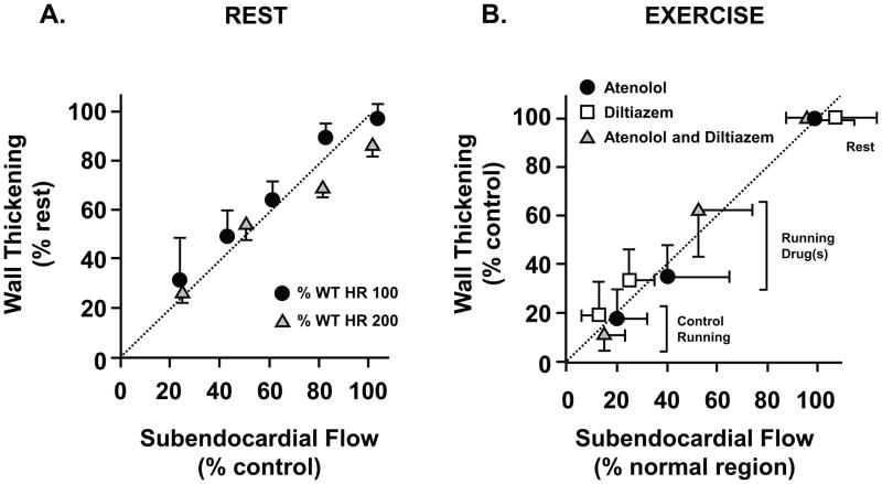
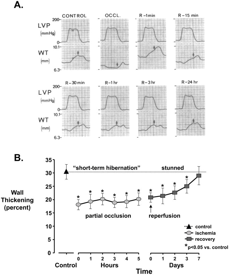
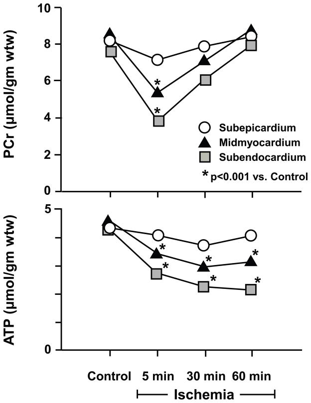
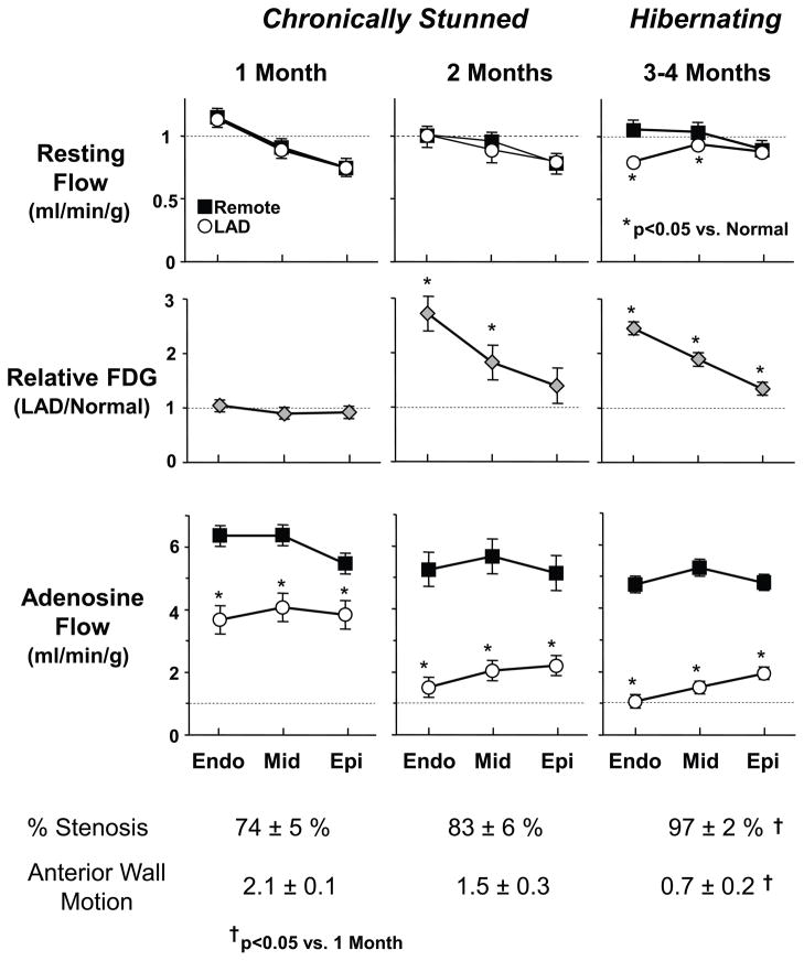
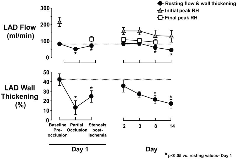
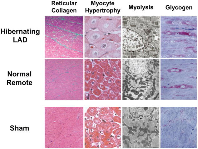
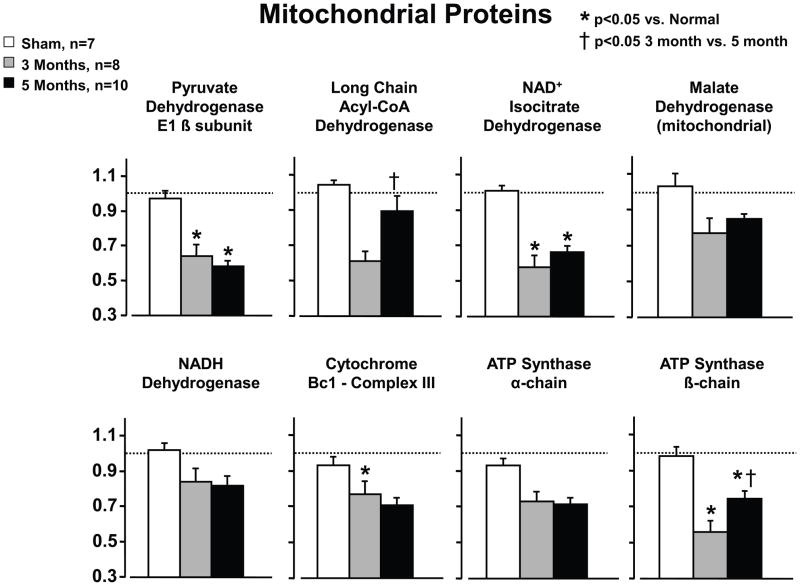
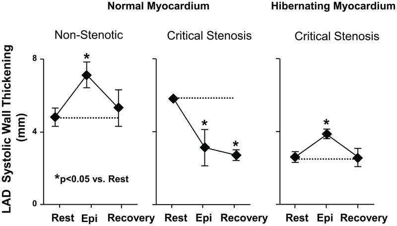
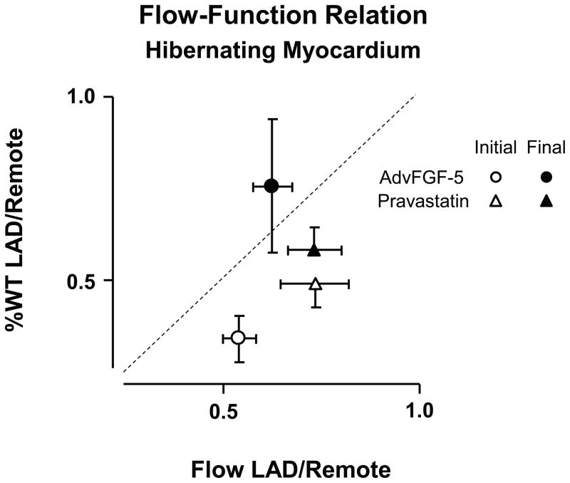
A large body of evidence has demonstrated that there is a close coupling between regional myocardial perfusion and contractile function. When ischemia is mild, this can result in the development of a new balance between supply and energy utilization that allows the heart to adapt for a period of hours over which myocardial viability can be maintained, a phenomenon known as "short-term hibernation". Upon reperfusion after reversible ischemia, regional myocardial function remains depressed. The "stunned myocardium" recovers spontaneously over a period of hours to days. The situation in myocardium subjected to chronic repetitive ischemia is more complex. Chronic dysfunction can initially reflect repetitive stunning with insufficient time for the heart to recover between episodes of spontaneous ischemia. As the frequency and/or severity of ischemia increases, the heart undergoes a series of adaptations which downregulate metabolism to maintain myocyte viability at the expense of contractile function. The resulting "hibernating myocardium" develops regional myocyte cellular hypertrophy as a compensatory response to ischemia-induced apoptosis along with a series of molecular adaptations that while regional, are similar to global changes found in advanced heart failure. As a result, flow-function relations become independently affected by tissue remodeling and interventions that stimulate myocyte regeneration. Similarly, chronic vascular remodeling may alter flow regulation in a fashion that increases myocardial vulnerability to ischemia. Here we review our current understanding of myocardial flow-function relations during acute ischemia in normal myocardium and highlight newly identified complexities in their interpretation in viable chronically dysfunctional myocardium with myocyte cellular and molecular remodeling. This article is part of a Special Issue entitled "Coronary Blood Flow".


Figures

Figure 1
Figure 1. Perfusion contraction matching during acute ischemia in normal myocardium
Relative reductions in function (wall thickening) are proportional to the relative reduction in subendocardial flow measured with microspheres in conscious dogs. This relationship is maintained during steady-state increases in myocardial work load over a wide range of heart rates during autoregulation (A) as well as during exercise with a fixed coronary stenosis (B). Medical interventions that ameliorate ischemia improve both subendocardial flow and wall thickening during exercise. HR - heart rate. (A modified from Canty et al [14, 17]; B modified from Matsuzaki et al. [62])
Figure 2
Figure 2. Stunned myocardium
(A) Myocardial stunning following a 15-minute total occlusion (OCCL). Wall thickening (WT) measured by ultrasonic crystals becomes dyskinetic, with systolic thinning. Upon reperfusion (R), function slowly improves and is completely normal after 24-hours. B, Myocardial stunning following a prolonged partial occlusion. During acute ischemia (circles), there is “short-term hibernation” reflecting an acute match between reduced flow, wall thickening, and metabolism. With reperfusion (squares), wall thickening remains depressed and gradually returns to normal after 1 week. LVP - left ventricular pressure. (A modified from Heyndrickx, et al. [21]; B modified from Matsuzaki M, et al. [25])
Figure 3
Figure 3. Metabolic matching during short-term hibernation
During moderate ischemia, reductions in contractile function are accompanied by adaptations in myocardial energy metabolism. These are characterized by an initial fall in creatine phosphate (PCr) and ATP. The transmural metabolite changes are greatest in the subendocardium (squares) reflecting the transmural variations in flow. With persistent ischemia, metabolism stabilizes with a regeneration of creatine phosphate that prevents further ATP depletion. While not shown, tissue lactate is transiently increased but normalizes with persistent ischemia. Adapted from Pantely et al. [26], and republished with permission of the American Heart Association, Inc.
Figure 4
Figure 4. Physiological progression from chronically stunned to hibernating myocardium in swine
Transmural microsphere flow measurements from pigs instrumented with a progressive LAD stenosis at rest and during adenosine vasodilation are shown along with transmural variations in 18FDG uptake (fasting conditions) obtained by ex vivo counting. Angiographic stenosis severity and anterior wall motion score (3 normal, 2 mild hypokinesis, 1 severe hypokinesis) at each time point are summarized below the graphs. As stenosis severity increases over time, vasodilated flow (adenosine) to the LAD region progressively falls, reflecting a functionally significant coronary lesion. Initially (1- and 2-months after instrumentation), anterior hypokinesis develops with normal resting flow consistent with chronically stunned myocardium. After 3-months, there is total occlusion with collateral-dependent myocardium and a critical impairment in coronary flow reserve. At this time, resting flow to the inner 2/3 of the LAD myocardium becomes reduced as compared to normally perfused remote myocardium and consistent with hibernating myocardium. The progression of abnormalities demonstrates that chronic stunning precedes the development of hibernating myocardium. In contrast to short-term hibernation resulting from primary reductions in coronary flow, the reduction in resting flow is a consequence, rather than cause of the contractile dysfunction in chronic hibernating myocardium (modified from Fallavollita et al. [39] with permission of the American Heart Association, Inc.).
Figure 5
Figure 5. Accelerated progression from stunned to hibernating myocardium by limiting reperfusion with a critical stenosis after a period of short-term hibernation in swine
Chronically instrumented swine were subjected to moderate ischemia for 15-minutes. In contrast to full reperfusion, regional LAD wall-thickening remained depressed after 24-hours when the coronary reactive hyperemic (RH) response was limited. When a acute critical stenosis was maintained during the subsequent 2-weeks, there was a progressive decline in function that was followed by a reduction in resting flow. The physiological and molecular changes were similar to pigs with hibernating myocardium that developed over 3-months. These results indicate that the physiological significance of a coronary stenosis (and spontaneous ischemia) rather than time is the major determinant of the progression from chronically stunned to hibernating myocardium. Moreover, this is accelerated by an abrupt increase in stenosis severity similar to that seen in patients presenting with an acute coronary syndrome. Republished from Thomas, et al. [24].
Figure 6
Figure 6. Myocyte cellular changes in swine with hibernating myocardium in the absence of heart failure
Swine with hibernating myocardium from a chronic LAD occlusion and collateral-dependent myocardium without infarction or heart failure develop an adapted phenotype. Like findings in humans, there is increased reticular connective tissue which is only ~2% greater than values in the remote normally perfused region (left column). While hearts appear grossly normal there is myocyte cellular hypertrophy in the hibernating region (second column) which reflects the consequences of apoptosis induced myocyte loss. The electron microscopic characteristics of hibernating myocardium are similar to humans with an adapted phenotype and demonstrate myofibrillar loss (Myolysis), numerous small mitochondria and increased glycogen content (right column). Interestingly, biopsies of remote, normally perfused segments from animals with an LAD occlusion show similar morphological changes. These data indicate that the “cellular hibernating phenotype” is not directly related to ischemia nor is it the cause of regional contractile dysfunction. Adapted from Canty [1].
Figure 7
Figure 7. Mitochondrial protein remodeling in animals with persistent hibernating myocardium
Proteomic profiling of animals with hibernating myocardium demonstrate a downregulation in mitochondrial proteins including some of the entry points to oxidative metabolism and selected components of the electron transport chain. These changes do not reflect mitochondrial loss or increased connective tissue. Like the physiological changes, most of the mitochondrial proteomic changes persisted in animals with hibernating myocardium that were studied 5-months after instrumentation. Exceptions included long chain Acyl-CoA dehydrogenase and the ATP synthase-β-chain which tended to normalize. While not shown, increases in stress proteins remained persistently elevated but increases in cytoskeletal proteins normalized in animals with longer term hibernation (5-months). Adapted from Page et al. [51] and republished with permission of the American Heart Association.
Figure 8
Figure 8. Protection of hibernating myocardium from acute demand-induced stunning following transient increases in demand during epinephrine infusion
LAD systolic wall thickening in animals with a critical stenosis and hibernating myocardium (n=9) were compared to normal myocardium subjected to an acute flow-limiting stenosis (n=3) on the LAD and non-stenotic controls (n=7). Hemodynamic changes were similar among groups. While LAD wall thickening was depressed in pigs with hibernating myocardium (right graph), it increased during β-adrenergic stimulation and returned to the identical depressed baseline level 15-minutes into recovery. The same transient increase and return to normal was also found in shams not subjected to acute ischemia (left graph). In contrast, animals instrumented with an acute critical stenosis that prevented flow from increasing during vasodilation developed contractile dysfunction during epinephrine induced increases in demand indicative of acute ischemia. In addition, in normal myocardium regional function remained depressed 15-minutes into recovery indicative of acute stunning. These results indicate that pigs with chronic hibernating myocardium develop protection against acute myocardial stunning. Adapted from Hu, et al. [50] and reprinted with permission.
Figure 9
Figure 9. Variability of Myocardial Flow-function relations in the absence of increased coronary flow in pigs with chronic hibernating myocardium
Relative wall thickening reductions in hibernating LAD regions vs. remote myocardium (%LADWT/%RemoteWT) vs. the relative reduction in resting subendocardial flow (LAD/Remote). The dashed line indicates a 1:1 relation between subendocardial flow and wall thickening reductions. In hibernating LAD regions, resting function was reduced more than subendocardial flow. Both adenoviral gene transfer of FGF-5 (fibroblast growth factor 5) and the HMG-Co-A reductase inhibitor pravastatin increased function with no change in resting flow or change in flow during pharmacological vasodilation (data not shown). These functional changes were accompanied by evidence of myocyte proliferation as reflected by Ki67 staining and, in the case of pravastatin, an increase in myocyte nuclear density. These data demonstrate alterations in the resting flow-function relation in hibernating myocardium that are accompanied by interventions that alter myocyte proliferation but have no effect on resting or maximal perfusion. (Data from Suzuki, et al. [52, 53]).
Figure 10
Figure 10. Structural and Functional remodeling of coronary resistance arterioles in hibernating myocardium
Panels A, B and C depict mild, moderate and severe vascular wall hypertrophy from arterioles in chronically instrumented swine subjected to 4-weeks of a severe coronary artery stenosis (Modified from Hong, et al. [58]). Lower graphs demonstrate arteriolar functional responses in vessels isolated from swine with chronic hibernating myocardium (Modified from Sorop, et al. [59]). While endothelium-dependent bradykinin-induced arteriolar vasodilation and passive vasodilation to nitroprusside (SNP) were normal (panels D and E), the constrictor response to endothelin was accentuated in vessels isolated from pigs with hibernating myocardium. This was also accompanied by an increase in circulating endothelin levels and an attenuated myogenic response in vitro. Collectively, these results demonstrate that viable chronically dysfunctional myocardium may have alterations in resistance artery control that may increase vascular tone and minimal coronary resistance. Modified and republished with permission of the American Heart Association.

References

    1. Canty JM., Jr . Coronary blood flow and myocardial ischemia. In: Bonow RO, Mann DL, Zipes DP, Libby P, editors. Braunwald’s Heart Disease. 9. Philadelphia: Elsevier; 2012. pp. 1049–75.
    1. Ross J., Jr Myocardial perfusion-contraction matching: Implications for coronary heart disease and hibernation. Circulation. 1991;83:1076–83. - PubMed
    1. Kloner RA, Jennings RB. Consequences of brief ischemia: stunning, preconditioning, and their clinical implications: part 1. Circulation. 2001;104:2981–9. - PubMed
    1. Bolli R. Myocardial ‘stunning’ in man. Circulation. 1992;86:1671–91. - PubMed
    1. Braunwald E, Kloner RA. The stunned myocardium: Prolonged, postischemic ventricular dysfunction. Circulation. 1982;66:1146–9. - PubMed

Publication types