. 2011 Nov 1;17(21):6712-22.
doi: 10.1158/1078-0432.CCR-11-0968. Epub 2011 Sep 2.

Species D adenoviruses as oncolytics against B-cell cancers

Affiliations

Species D adenoviruses as oncolytics against B-cell cancers

Christopher Y Chen et al. Clin Cancer Res. .

Abstract

Purpose: Oncolytic viruses are self-amplifying anticancer agents that make use of the natural ability of viruses to kill cells. Adenovirus serotype 5 (Ad5) has been extensively tested against solid cancers, but less so against B-cell cancers because these cells do not generally express the coxsackie and adenoviral receptor (CAR). To determine whether other adenoviruses might have better potency, we "mined" the adenovirus virome of 55 serotypes for viruses that could kill B-cell cancers.

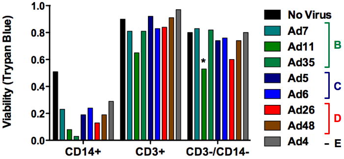
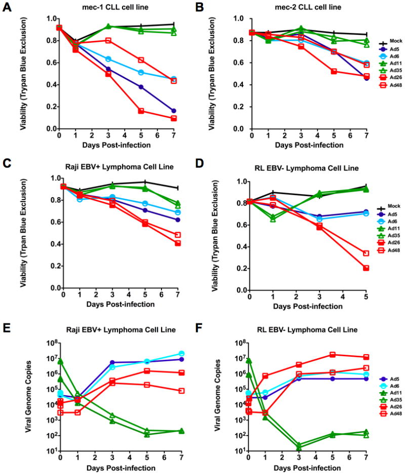
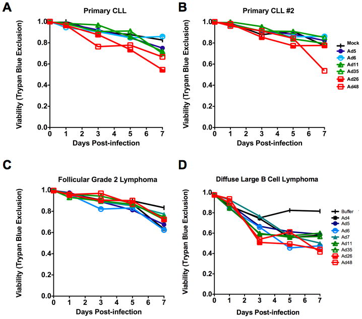
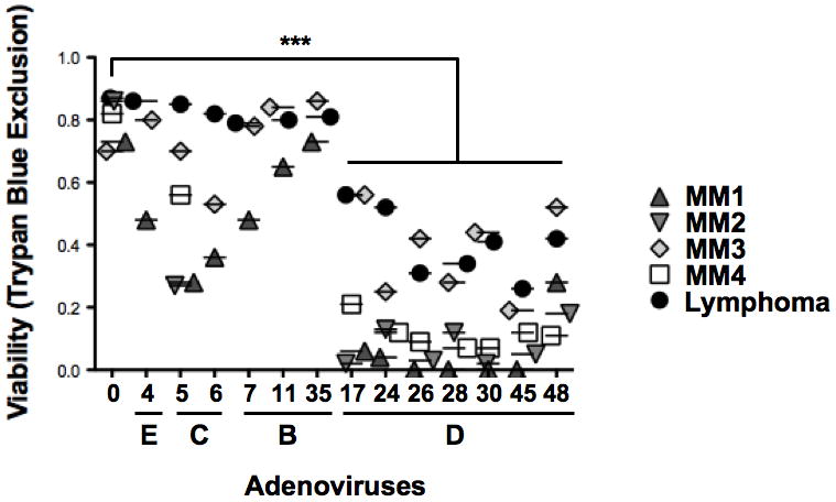
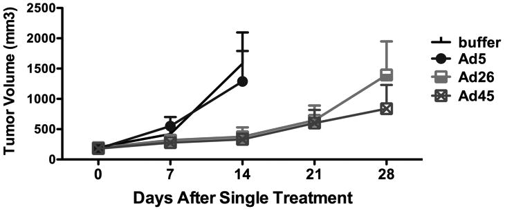
Experimental design: Fifteen adenoviruses selected to represent Ad species B, C, D, E, and F were tested in vitro against cell lines and primary patient B-cell cancers for their ability to infect, replicate in, and kill these cells. Select viruses were also tested against B-cell cancer xenografts in immunodeficient mice.

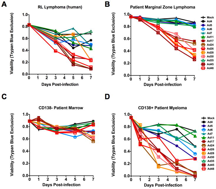
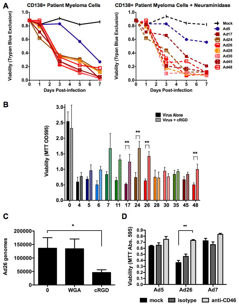
Results: Species D adenoviruses mediated most robust killing against a range of B-cell cancer cell lines, against primary patient marginal zone lymphoma cells, and against primary patient CD138+ myeloma cells in vitro. When injected into xenografts in vivo, single treatment with select species D viruses Ad26 and Ad45 delayed lymphoma growth.

Conclusions: Relatively unstudied species D adenoviruses have a unique ability to infect and replicate in B-cell cancers as compared with other adenovirus species. These data suggest these viruses have unique biology in B cells and support translation of novel species D adenoviruses as oncolytics against B-cell cancers.


Conflict of interest statement

Conflicts of Interest: None

Figures

Figure 1
Figure 1. Comparison of adenoviral serotypes for infection and killing of normal PBMCs
The indicated cells were incubated for seven days with the indicated wild-type adenoviruses and cell viability was assessed by Trypan Blue exclusion. MOI = 10,000 viral particles/cell.
Figure 2
Figure 2. Comparison of adenoviral serotypes for infection and replication in B-cell cancer cell lines
Comparison of cell killing of either MEC1 (A) or MEC2 (B) chronic lymphocytic leukemia cell lines by adenoviruses as assessed by Trypan Blue uptake following infection with virus at an MOI of 10,000 viral particles/cell. Killing of either Raji (C) or RL (D) human lymphoma cell lines by adenoviruses as assessed by Trypan Blue uptake. E and F. Real-time quantitative PCR showing viral genome copies present within cells over a one week time course following infection for cells in C and D. Species B (green), species C (blue), or species D (yellow and red). N=3.
Figure 3
Figure 3. Comparison of adenoviral serotypes for infection and replication of primary B cell cancer cells
A and B. One week cell viability of two different CLL patient’s sample cells after infection with six different adenoviral serotypes. C. Viability of follicular grade 2 cancer lymphoma patient sample cells following infection by a panel of eight adenoviral serotypes. D. Viability of diffuse large B cell lymphoma patient sample cells following infection by panel of eight adenoviral serotypes. Experiments were performed using a viral MOI of 10,000 particles/cell.
Figure 4
Figure 4. Expanded comparison of adenoviral serotypes for infection and replication in B-cell cancer cells
A 13-member panel of adenoviral serotypes are tested against two B cell cancers. Lymphoma - (A) RL human lymphoma (B) Patient marginal zone lymphoma. Myeloma – (C) non-malignant CD138- patient bone marrow cells (D) malignant C138+ patient bone marrow cells. Viability was assessed by Trypan Blue uptake following infection at an MOI of 10,000 viral particles/cell. Green = species B; blue = species C; yellow, orange, red = species D, and brown = species E.
Figure 5
Figure 5. Pooled cell viability data from Five Patient B-cell cancers
The marginal zone lymphoma patient sample data was pooled with cell killing data of four patient multiple myeloma samples. Individual viabilities (y-axis) from each experiment are presented as points above the viral serotype tested (x-axis).
Figure 6
Figure 6. In vivo comparison of select adenoviral serotypes for killing of lymphoma xenografts in nude mice
RL lymphoma tumors were initiated subcutaneously in nude mice. Groups of six tumor-bearing mice were injected intratumorally a single time with 3×10¹⁰ virus particles (vp) of the indicated adenoviruses. Tumor size was monitored over time after injection and mean tumor sizes are shown. Lines end when the first animal of the group had to be sacrificed to avoid misleading skewing of the lines as larger tumors are removed.
Figure 7
Figure 7. Receptor blocking during species D adenovirus infection
A. 13-panel adenoviral infection (MOI=1,000 viral particles/cell) of patient CD138+ multiple myeloma cells with (right) and without (left) prior treatment with neuraminidase to remove sialic acid. B. Viability of Hep3B cells one week following infection by the 13 adenoviruses (x-axis) in the presence or absence of cyclic RGD peptide. “0” = uninfected. P-value of two-tailed T-test. C. Effects of wheat germ agglutinin (WGA) or cyclic RGD on Ad26 binding to Hep3B cells as assessed by qPCR. “0” = uninfected. P-value of one-tailed T-test. D. Blocking CD46 binding of Ad26 to human patient reactive follicular hyperplasia cells by addition of isotype or anti-CD46 antibody. * = p < 0.05. ** = p < 0.01.

References

    1. Altekruse SFKC, Krapcho M, Neyman N, Aminou R, Waldron W, Ruhl J, Howlader N, Tatalovich Z, Cho H, Mariotto A, Eisner MP, Lewis DR, Cronin K, Chen HS, Feuer EJ, Stinchcomb DG, Edwards BK. SEER Cancer Statistics Review. 2010. National Cancer Institute; Apr 15, 2010.
    1. Jemal A, Siegel R, Ward E, Hao Y, Xu J, Murray T, et al. Cancer statistics, 2008. CA Cancer J Clin. 2008;58:71–96. - PubMed
    1. Cattaneo R, Miest T, Shashkova EV, Barry MA. Reprogrammed viruses as cancer therapeutics: targeted, armed and shielded. Nat Rev Microbiol. 2008;6:529–40. - PMC - PubMed
    1. Barry MA, Hofherr SE, Chen CY, Senac JS, Hillestad ML, Shashkova EV. Systemic delivery of therapeutic viruses. Curr Opin Mol Ther. 2009;11:411–20. - PubMed
    1. Shayakhmetov DM, Papayannopoulou T, Stamatoyannopoulos G, Lieber A. Efficient gene transfer into human CD34(+) cells by a retargeted adenovirus vector. J Virol. 2000;74:2567–83. - PMC - PubMed

Publication types

MeSH terms