. 2011 Dec 1;20(23):4617-33.
doi: 10.1093/hmg/ddr396. Epub 2011 Sep 1.

A valid mouse model of AGRIN-associated congenital myasthenic syndrome

Affiliations

A valid mouse model of AGRIN-associated congenital myasthenic syndrome

Laurent P Bogdanik et al. Hum Mol Genet. .

Abstract

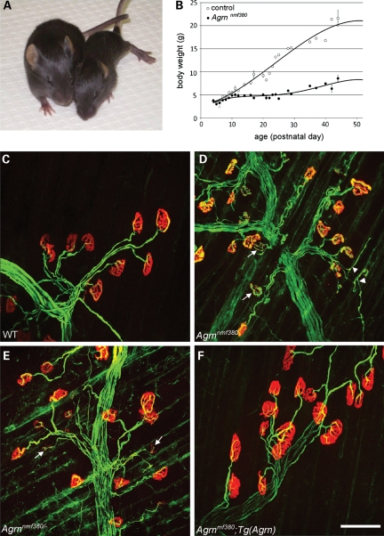
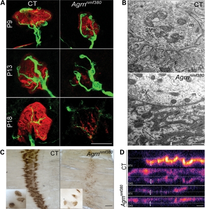
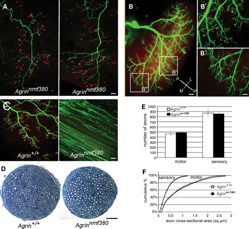
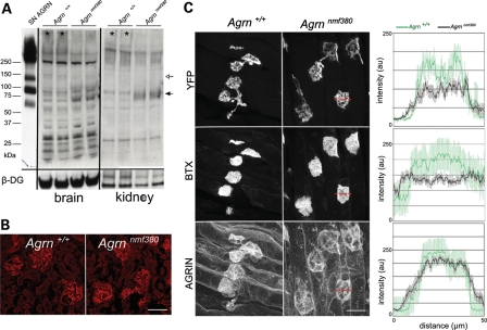
Congenital myasthenic syndromes (CMS) are inherited diseases affecting the neuromuscular junction (NMJ). Mutations in AGRIN (AGRN) and other genes in the AGRIN signaling pathway cause CMS, and gene targeting studies in mice confirm the importance of this pathway for NMJ formation. However, these mouse mutations are complete loss-of-function alleles that result in an embryonic failure of NMJ formation, and homozygous mice do not survive postpartum. Therefore, mouse models of AGRIN-related CMS that would allow preclinical testing or studies of postnatal disease progression are lacking. Using chemical mutagenesis in mice, we identified a point mutation in Agrn that results in a partial loss-of-function allele, creating a valid model of CMS. The mutation changes phenylalanine 1061 to serine in the SEA domain of AGRIN, a poorly characterized motif shared by other extracellular proteoglycans. NMJs in homozygous mice progressively degrade postnataly. Severity differs with genetic background, in different muscles, and in different regions within a muscle in a pattern matching mouse models of motor neuron disease. Mutant NMJs have decreased acetylcholine receptor density and an increased subsynaptic reticulum, evident by electron microscopy. Synapses eventually denervate and the muscles atrophy. Molecularly, several factors contribute to the partial loss of AGRIN's function. The mutant protein is found at NMJs, but is processed differently than wild-type, with decreased glycosylation, changes in sensitivity to the protease neurotrypsin and other proteolysis, and less efficient externalization and secretion. Therefore, the Agrn point mutation is a model for CMS caused by Agrn mutations and potentially other related neuromuscular diseases.


Figures

Figure 1.
Figure 1.
Overt phenotype and NMJ disaggregation in a new Agrn allele. (A) By P15, affected mice (right) are significantly smaller than their WT littermates, display altered locomotion and signs of muscle stiffness and atrophy. (B) Growth curves of mutant and unaffected littermates. Although the bodyweight is the same at birth, significant differences are observed by 2 weeks of age and by P21, affected mice weigh approximately half as much as controls. NMJs in control (C) and mutant (D) mice at P15. In control mice, the motor nerve terminal (green, labeled with anti-neurofilament and anti-SV2) completely overlaps the postsynaptic AChRs (red, labeled with α-BTX) on the muscle and the synapse is assuming its mature morphology. In mutant mice, the synapse still resembles an immature plaque, AChR staining intensity is reduced, AChR clusters begin to disaggregate (arrows) and motor nerve terminals are beginning to sprout beyond the endplates (arrowheads). (E) In genetic complementation tests, the nmf380 allele failed to complement Agrn knockout alleles, with similar overt and NMJ phenotypes (arrows) to nmf380 homozygotes. (F) The phenotype was completely rescued by an Agrn genomic transgene. Scale bar in (F) is 100 µm.
Figure 2.
Figure 2.
Molecular characterization of the new Agrn mutation. (A) Sequencing of cDNA from Agrnnmf380 homozygotes reveals an A to G change in exon 18 of Agrn, resulting in a change of phenylalanine 1061 to serine (F1061S) in the protein. (B) F1061 (*) is in the SEA module of AGRIN, a poorly characterized domain in the middle of the AGRIN protein. Other domains include the type II transmembrane SN-N-terminus, the secreted, laminin-binding LN-N-terminus, folistatin repeats (F), sites of glycosaminoglycan addition (GAG), laminin domain (L), serine/threonine-rich repeats (ST), epidermal growth factor like repeats (EGF1-4), laminin-type globular domains (G1–3) and sites of alternative splicing (X, Y, Z). (C) A phenylalanine at this position in AGRIN (dark blue highlight) is conserved in rodents, human, chick, zebrafish (danre) and electric ray (disom), as well as in the SEA modules of mouse enterokinase, mucins 1, 13 and 16 (Q9D1H1). (D) A structural prediction of the SEA domain of AGRIN extrapolated from the crystal structure of Mucin 16 shows the mutation changes a phenylalanine located at the carboxy terminal end of the first alpha-helix of the SEA module (arrowhead). The autocatalytic cleavage site found in the mucins in a hinge region is also indicated (arrow). (E) Analysis of mRNA levels by northern blotting indicates that there is no reduction in Agrn transcripts or changes in transcript size in Agrnnmf380 homozygotes, as anticipated for a point mutation. Control (WT) and mutant (M) lanes are indicated, β-Actin was used as a loading control. R) Reverse-transcription PCR on brain cDNA from control (WT) and mutant (M) mice with primers in exons 31 and 34, flanking the alternative Z-splicing site (exons 32 and 33). DNA molecular weight standard is on the left side and PCRs on cloned Z0, Z8 and Z19 cDNAs with the same primers are shown on the right. Alternative splicing at the Z-splicing site is critical for AGRIN's activity and splicing at this site is not changed by the mutation.
Figure 3.
Figure 3.
The F1061S mutation causes a postnatal onset NMJ disaggregation. (A) Time course of the NMJ formation and disaggregation on the triangularis sterni muscle. Labeling with BTX (red), and Thy-1-YFP16 (green) demonstrates a progressive defects in both pre- and postsynaptic features of the NMJ. Scale bar 25 μm. (B) Transmission electron microscopy on the plantaris muscle at P20. Hallmarks of presynaptic (mitochondria, M, synaptic vesicles, SV) and postsynaptic (folds, F) differentiation are clear in control NMJs, and are present but abnormal in mutant NMJs, suggesting the synapses begin to mature, forming postsynaptic folds for example, and then disperse. Scale bar 500 nm. (C) ECM-bound AChE is also removed from the NMJs in the mutant, as demonstrated by a histochemical stain for AChE activity, although an endplate band is still discernable. Scale bar is 500 μm (50 μm for the inserts). (D) Beta-dystroglycan is also disrupted at the NMJ. Confocal stacks where the BTX (blue, top) and beta-dystroglycan (DAG, red, bottom) channels are separated, and viewed in the X–Z plane in false color. The AChR labeling colocalizes with synaptic dystroglycan (brackets) whose intensity is reduced in Agrnnmf380 homozygotes. Scale bar 5 μm.
Figure 4.
Figure 4.
Variability of the NMJ phenotype between muscles and transition to a slow-fiber phenotype. (A) Comparison of the NMJ disaggregation in the diaphragm (Dia), a muscle affected early, and the skin-facing region of the TA, a muscle affected later at postnatal days (P) 1, 4 and 14. Presynaptic overgrowth and decreased intensity of AChR staining is evident in the diaphragm at P1, but is not evident in the TA until later ages. Scale bar 100 μm. (B) Quantification of the disaggregation of the NMJs in three representative muscles, the diaphragm, TA and soleus (Sol). Bars indicate the AChR cluster area, lines indicate the intensity of BTX labeling of AChRs expressed as a percentage of control values. (C) The NMJs of extraocular muscles are preserved at P30, an age where NMJs are severely affected in all other muscles. Scale bar 100 μm. (D) Type I, slow-myosin staining (green, BTX in red) in cross-sections of the lateral gastrocnemius at P20 demonstrates an increase in slow fibers in the Argnnmf380 mutant mice. Scale bar 50 μm.
Figure 5.
Figure 5.
Agrnnmf380 shares features with motor-neuron disease models, but is not a die-back neuropathy. (A) Two individual motor units in the triangularis sterni at P10, visualized with the Thy1.1-YFP-H transgenic strain in Agrnnmf380/nmf380 mice (green). Left, all the terminals of the motor unit are still connected to their NMJs (α-BTX, red). Right, all the terminals of the motor unit have lost their connections and are sprouting, indicating that preservation or disaggregation of NMJs is synchronous at the level of motor units. (B) Innervation of the lateral gastrocnemius of P10 mutants visualized with transgenic Thy1-YFP16. The antero-posterior and medio-lateral axes are shown in the lower right corner. Higher magnification of the intermediate (B′) and medial (B″) regions is shown on the right. Most of NMJs of the intermediate region (B′) are still innervated and axon terminals facing disaggregated NMJs do not extend beyond the postsynaptic site. In the medial region (B″), most of NMJs are disaggregated and axon terminals are abundantly sprouting. (C) Medial aspect of the quadriceps in 3-month-old control and mutant mice. NMJs have disaggregated in the mutant muscle and presynaptic terminals are sprouting throughout the muscle. (D) Sections of the motor branch of the femoral nerve innervating the quadriceps in 3-month-old mice show neither a noticeable loss of axons nor a reduction in their diameter in the mutant nerves. (E and F) Quantification of the number of axons in the motor and sensory branches of the femoral nerve (E) and a cumulative histogram of their cross-sectional areas (F) at 3 months of age show no significant axon loss or atrophy, more than 2 months after the loss of most of NMJs in the quadriceps (n = 4 controls and 4 mutants). Scale bars: 100 μm in A, B′, B″ and C; 200 μm in (B) and 50 μm in (D).
Figure 6.
Figure 6.
Genetic background effects in Agrn mutant mice. (A) Muscle weight (mg): body weight (g) ratios illustrate the neurogenic atrophy of the lateral and medial gastrocnemius (LGC and MGC), TA and plantaris (Pl) in P15–P30 Agrnnmf380/nmf380 mutants in a C57BL6 or DBA/2 background (pooled measures on two controls and eight mutants). In an N3.CAST background (3 mutants, 3 controls, 10 months old), the atrophy of these muscles was less severe and not significant. Student t-test, *P < 0.05, **P < 0.01. Inset, body weights are reduced in mutant animals relative to age-matched littermates in a C57BL/6 background (P20), but not in a CAST background (10 months). (B) NMJs of the TA display an abnormal shape but are still partially innervated in 10-month-old mutants in an N3 CAST background. Scale bar 10 μm. (C) The endplate band of the TA shown by BTX staining. Genotypes are indicated at the bottom of (D). C57BL/6 animals were 1 month old, N3 CAST animals were 10 months old. Scale bar 100 μm. (D) Hematoxylin and eosin staining on hindlimb cross-sections shows the developmental delay and atrophy of all mutant muscles in the C57BL/6 background, whereas the atrophy is very limited in the N3.CAST background. Inserts show high magnification of the muscle fibers. While most of the fibers have a polygonal shape and are tightly arranged in the controls (Agrn+/+), numerous rounded, smaller fibers characteristic of a neurogenic atrophy, are seen in the mutant in a C57BL/6 background. A few rounded fibers are also seen in the N3 CAST mutant (asterisk), but the atrophy is limited. C57BL/6 animals were 1 month old, N3 CAST animals were 10 months old. Scale bar is 1 mm on the top panels and 100 μm in the inserts.
Figure 7.
Figure 7.
AGRIN abundance and deposition in tissues. (A) Western blot on brain and kidney protein preparations. Using an antibody that recognizes the C-terminus of AGRIN, glycosylated, full-length AGRIN (>250 kDa, *) is reduced in P30 mutant samples, and a shorter peptide (75 kDa, black arrow) is increased. Recombinant SN-AGRIN was run in parallel on the same gel and shows the full-length protein, the 110 kDa, NT-dependent cleavage product (white arrow) and the 75 kDa product. Beta-dystroglycan was used as a loading control. (B) LN-AGRIN deposition in P10 kidney glomeruli is normal in the mutants, as detected with an LN-specific N-terminal antibody. Scale bar 10 mm. (C) Quantitative immunofluorescence for agrin at the NMJs on P4 triangularis sterni. Representative images of the three separate channels (presynaptic Thy1-YFP, BTX and AGRIN) are shown. Measures of staining intensity (0–250) were made along a line across the NMJ profile (red lines) and averaged (right graphs, mean ± SEM). Scale bar 10 mm.
Figure 8.
Figure 8.
Glycosylation, NT cleavage and plasma membrane targeting are affected by the F1061S mutation. (A) Schematic of the expression constructs used. Full-length SN-AGRIN with C-terminal myc tag, C110 AGRIN with vector signal peptide and C-terminal myc tag, AGRIN-SEA domain fused to alkaline phosphatase with signal peptide and C-terminal myc tag, and full-length NT with HA tag. The NT cleavage sites in AGRIN, upstream of the SEA domain and upstream of the third G-domain are indicated on the SN-AGRIN schematic. (B) Western blot of transfected HEK293 cell lysates and their conditioned media. The western blot membranes were probed with either an anti-myc antibody to visualize the carboxy terminal myc tag on AGRIN, an anti-HA antibody to detect NT and anti-tubulin as a loading control. In cell membranes, full-length AGRIN is present in both wild-type and F1061S transfected cells, but the larger, glycosylated forms are reduced in the mutant protein. In conditioned media, cells transfected with wild-type AGRIN produce 110 and 29 kDa C-terminal fragments as a result of NT cleavage, as well as a non-NT-dependent 75 kDa product. In cells transfected with F1061S constructs, less NT cleavage product is seen without NT cotransfection, and only the C-terminal 29 kDa fragment is detected when NT is cotransfected, suggesting the N-terminal site near the SEA domain is insensitive to NT cleavage. (C) Live cell staining without fixation or permeabilization for the C-terminal myc tag on full-length, type II transmembrane AGRIN. Cells were co-transfected with a mitochondrial COX8-YFP fusion (green, to identify individual transfected cells), and WT or mutant AGRIN, and without or with NT. Anti-myc (red) detects externalized AGRIN in both wild-type and F1061S transfected cells, and this cell surface staining is lost with cotransfection with NT, indicating both proteins are sensitive to NT cleavage. (D) Western blots on transfected CHO cells and their conditioned media. Cells were transfected with the full-length, C110 or SEA-alkaline phosphatase fusion DNAs, with or without the F1061S mutation. Blots of cell lysates and media were probed for the C-terminal myc tag. Full-length transmembrane AGRIN remains cell associated, whereas C110 AGRIN is secreted into the media in both wild-type and F1061S constructs. The F1061S SEA domain causes almost complete failure of secretion of the alkaline phosphatase fusion construct.

Similar articles

Cited by

References

    1. Engel A.G., Ohno K., Sine S.M. Chapter 66: Congenital Myasthenic Syndrome. In: Engel A.G., Franzini-Armstrong C., editors. Myology. 3rd edn. Vol. 2. New York: McGraw-Hill; 2004. pp. 1801–1854.
    1. Sanes J.R., Lichtman J.W. Development of the vertebrate neuromuscular junction. Ann. Rev. Neurosci. 1999;22:389–442. doi:10.1146/annurev.neuro.22.1.389. - DOI - PubMed
    1. Burden S.J. Building the vertebrate neuromuscular synapse. J. Neurobiol. 2002;53:501–511. doi:10.1002/neu.10137. - DOI - PubMed
    1. Kummer T.T., Misgeld T., Sanes J.R. Assembly of the postsynaptic membrane at the neuromuscular junction: paradigm lost. Curr. Opin. Neurobiol. 2006;16:74–82. doi:10.1016/j.conb.2005.12.003. - DOI - PubMed
    1. Nitkin R.M., Smith M.A., Magill C., Fallon J.R., Yao Y.M., Wallace B.G., McMahan U.J. Identification of agrin, a synaptic organizing protein from Torpedo electric organ. J. Cell Biol. 1987;105:2471–2478. doi:10.1083/jcb.105.6.2471. - DOI - PMC - PubMed

Publication types

MeSH terms