Clinical Trial
. 2011 Nov 3;118(18):4957-62.
doi: 10.1182/blood-2011-07-368084. Epub 2011 Sep 2.

The CXCR4 antagonist plerixafor corrects panleukopenia in patients with WHIM syndrome

Affiliations
Clinical Trial

The CXCR4 antagonist plerixafor corrects panleukopenia in patients with WHIM syndrome

David H McDermott et al. Blood. .

Abstract

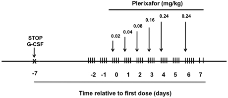
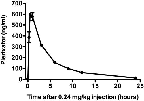
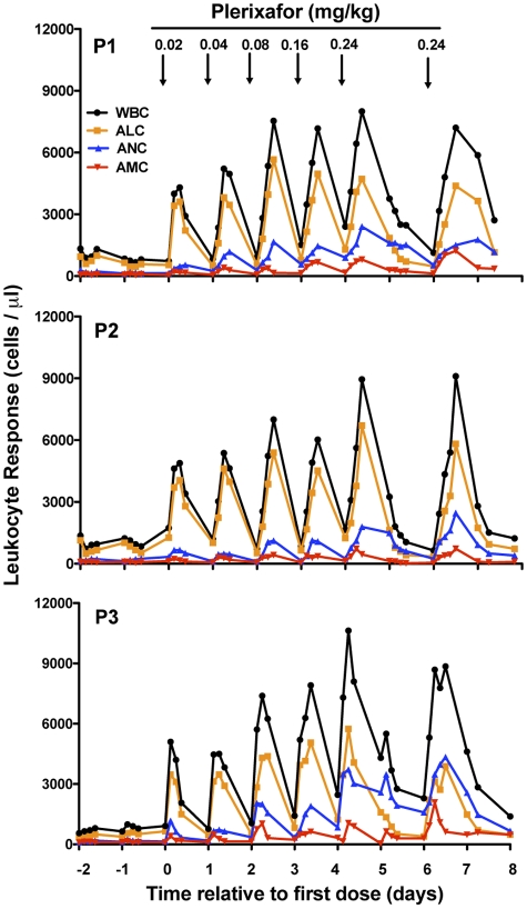
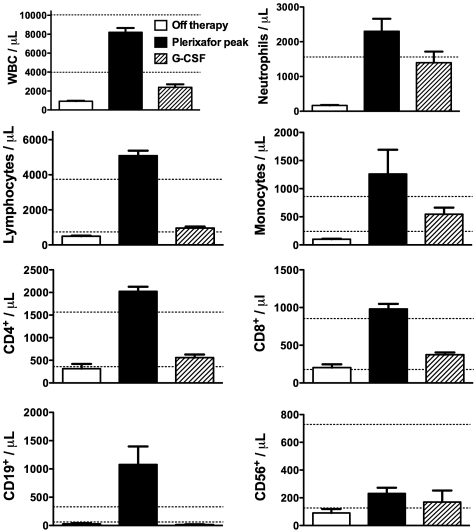
WHIM syndrome is a rare congenital immunodeficiency disorder characterized by warts, hypogammaglobulinemia, infections, and myelokathexis (neutropenia because of impaired egress from the BM); most patients also have severe panleukopenia. Because WHIM syndrome is caused by mutations in the chemokine receptor CXCR4 that result in increased agonist-dependent signaling, we hypothesized that the CXCR4 antagonist plerixafor (Mozobil [Genyzme Corporation], AMD3100), might be an effective treatment. To test this, we enrolled 3 unrelated adult patients with the most common WHIM mutation, CXCR4(R334X), in a phase 1 dose-escalation study. Plerixafor increased absolute lymphocyte, monocyte, and neutrophil counts in blood to normal without significant side effects in all 3 patients. Peak responses occurred at 3-12 hours after injection and waned by 24 hours after injection which tracked the drug's pharmacokinetics. All 3 cell types increased in a dose-dependent manner with the rank order of responsiveness absolute lymphocyte > monocyte > neutrophil. These data provide the first pharmacologic evidence that panleukopenia in WHIM syndrome is caused by CXCL12-CXCR4 signaling-dependent leukocyte sequestration, and support continued study of plerixafor as mechanism-based therapy in this disease. This study is registered at http://www.clinicaltrials.gov as NCT00967785.


Figures

Figure 1
Figure 1
Design of a phase 1 study of plerixafor treatment in adults with WHIM syndrome. Patients receiving G-CSF had the drug discontinued at the indicated time. Arrows indicate time of subcutaneous administration of plerixafor at the indicated dose. Vertical hash marks denote times of blood sampling for complete blood counts. Not shown: a sham saline injection of the same volume as the lowest drug dose was given on day −1 and the greatest drug dose on day +7.
Figure 2
Figure 2
Plerixafor pharmacokinetics in WHIM patients. A single dose of 0.24 mg/kg of plerixafor was subcutaneously injected on day +6 as indicated in Figure 1. Plasma drug concentration was determined at the times indicated. Shown are the mean ± SEM of data from patient 1 and 2.
Figure 3
Figure 3
Plerixafor normalizes absolute blood leukocyte counts in patients with WHIM syndrome. All data collected from all 3 patients studied (P1, P2, and P3) are shown. Each point represents a single value obtained from that time point. Time zero is defined as the first day of plerixafor administration. Arrows indicate the time when the indicated dose of plerixafor in mg/kg was administered subcutaneously to the patient (Figure 1). Not shown: a sham saline injection of the same volume as the lowest dose was given on day −1 and as the greatest dose on day +7. WBC indicates white blood count; ALC, absolute lymphocyte count; ANC, absolute neutrophil count, and AMC, absolute monocyte count.
Figure 4
Figure 4
Comparison of blood leukocyte counts in WHIM patients after treatment with plerixafor and G-CSF. Parameters on x-axis are defined as follows: (1) “Plerixafor peak,” the mean ± SEM of the peak absolute blood leukocyte subset counts of WHIM patients 1, 2, and 3 in response to administration of both 0.16 and 0.24 mg/kg doses of plerixafor in this study; (2) “Off therapy,” the mean ± SEM of absolute blood leukocyte subset counts for all values determined on days −1 and −2 of the phase 1 study (Figure 1); and (3) “G-CSF,” the mean ± SEM of all available absolute blood leukocyte subset counts of WHIM patients 1 and 2 during treatment with daily G-CSF at 1-3 μg/kg. Horizontal dotted lines indicate the normal range.
Figure 5
Figure 5
Neutrophils released by plerixafor function normally. (A) ROS production. DHR was loaded into isolated neutrophils, which were then stimulated with PMA. Data are from a single experiment in which purified blood neutrophils from patient 2 were used, gated as shown in orange on the left and displayed as a histogram on the right. Data are representative of 2 independent experiments, one each for patients 2 and 3, each tested on day +4 of the protocol (Figure 1). Neutrophils were obtained 3 hours after the administration of 0.24 mg/kg plerixafor. FSC indicates forward scatter; SSC, side scatter. (B) Staphylocidal assay. S aureus was incubated at either 2 or 8 bacteria/neutrophil for the time indicated on the x-axis in vitro at 37°C. Cells were then lysed and plated on agar to determine the viable bacterial count. Data are from a single experiment using neutrophils from patient 2 and a single healthy donor (normal) incubated at 2 bacteria/cell and are representative of 4 experiments performed, one for patients 2 and 3 at the 2 concentrations of bacteria. Patient neutrophils were obtained on day +4 of the protocol, 3 hours after the administration of 0.24 mg/kg plerixafor (Figure 1). Control, S aureus incubated without neutrophils.

Comment in

  • Released on a WHIM.
    Diaz GA. Diaz GA. Blood. 2011 Nov 3;118(18):4764-5. doi: 10.1182/blood-2011-08-375162. Blood. 2011. PMID: 22053172 No abstract available.

References

    1. Zuelzer WW. “Myelokathexis”—a new form of chronic granulocytopenia. Report of a case. N Engl J Med. 1964;270:699–704. - PubMed
    1. Krill CE, Jr, Smith HD, Mauer AM. Chronic idiopathic granulocytopenia. N Engl J Med. 1964;270:973–979. - PubMed
    1. Wetzler M, Talpaz M, Kleinerman ES, et al. A new familial immunodeficiency disorder characterized by severe neutropenia, a defective marrow release mechanism, and hypogammaglobulinemia. Am J Med. 1990;89(5):663–672. - PubMed
    1. Kawai T, Malech HL. WHIM syndrome: congenital immune deficiency disease. Curr Opin Hematol. 2009;16(1):20–26. - PMC - PubMed
    1. Hernandez PA, Gorlin RJ, Lukens JN, et al. Mutations in the chemokine receptor gene CXCR4 are associated with WHIM syndrome, a combined immunodeficiency disease. Nat Genet. 2003;34(1):70–74. - PubMed

Publication types

MeSH terms

Supplementary concepts

Associated data