. 2011 Nov;301(5):H1897-906.
doi: 10.1152/ajpheart.00260.2011. Epub 2011 Sep 2.

Nitric oxide formation by lymphatic bulb and valves is a major regulatory component of lymphatic pumping

Affiliations

Nitric oxide formation by lymphatic bulb and valves is a major regulatory component of lymphatic pumping

H Glenn Bohlen et al. Am J Physiol Heart Circ Physiol. 2011 Nov.

Abstract

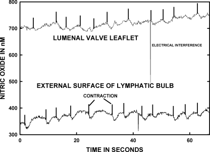
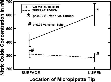
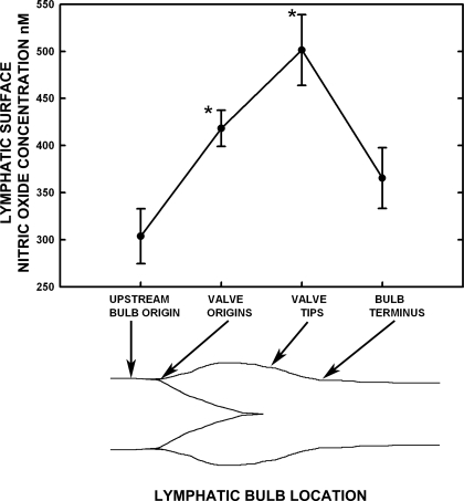
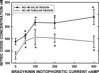
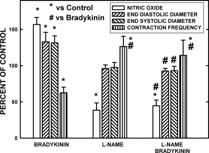
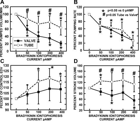
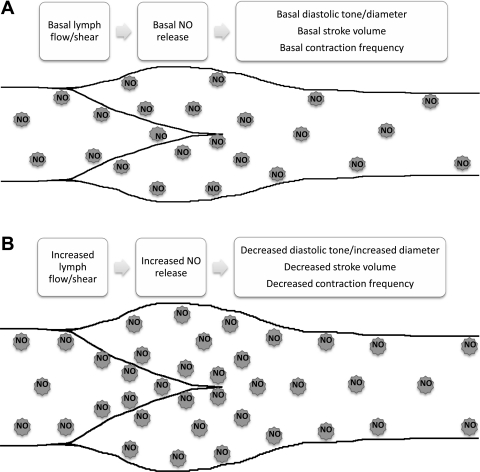
Microscopic lymphatics produce nitric oxide (NO) during contraction as flow shear activates the endothelial cells. The valve leaflets and bulbous valve housing contain a large amount of endothelial nitric oxide synthase (eNOS) due both to many endothelial cells and increased expression of eNOS. Direct NO measurements indicate the valve area has a 30-50% higher NO concentration ([NO]) than tubular regions although both regions generate equivalent relative increases in [NO] with each contraction. We hypothesize that 1) the greater eNOS and [NO] of the bulb region would have greater effects to lower pumping activity of the overall lymphatic than occurs in tubular regions and 2), the elevated [NO] in the bulb region may be because of high NO production in the valve leaflets that diffuses to the wall of the bulb. Measurement of [NO] with a micropipette inside the lymphatic bulb revealed the valve leaflets generate ~50% larger [NO] than the bulb wall in the in vivo rat mesenteric lymphatics. The valves add NO to the lymph that quickly diffuses to the bulb wall. Bradykinin locally released iontophoretically from a micropipette on both bulbs and tubes increased the [NO] in a dose-dependent manner up to ~50%, demonstrating agonist activation of the NO pathway. However, pumping output determined by contraction frequency and stroke volume decreased much more for the bulb than tubular areas in response to the bradykinin. In effect, NO generation by the bulb area and its valves limits the pumped flow of the total lymphatic by lowering frequency and stroke volume of individual contractions.


Figures

Fig. 1.
Fig. 1.
Nitric oxide concentration ([NO]) of the external bulb surface and internal valve leaflet are shown for the same lymphatic. The data were not obtained simultaneously but within 10 min of each other. When the nitric oxide (NO) micropipette is very near and essentially touching the valve leaflet surface, the recorded [NO] was consistently much higher than on the outer surface of the bulb wall. The vertical marks on each record indicate the onset of mechanical contraction by the lymphatic bulb followed by a transient decrease in [NO] as the valve closed and then an increase in [NO] presumably activated by the transient increase in lymph flow before the valve closed. The same mechanical correlation to changes in [NO] was more evident for the bulb external surface than in the lumen near the lymphatic valves.
Fig. 2.
Fig. 2.
The luminal and surface [NO] of lymphatic bulb and tubular regions are shown as averages for 11 bulb and 12 tube regions in 9 animals with dual bulb and downstream tubular measurements in all but one animal. The tubular region was ∼500 μm downstream from the terminal part of the lymphatic bulb. The valve lumen [NO] was consistently much higher than that of the bulb surface near the terminal portion of the valve leaflets. The tubular surface and luminal [NO] were very similar and within the resolution of the NO microelectrode technology in most cases. In all animals, the tubular wall [NO] was lower than that of the bulb surface. *Significant difference from the bulb origin.
Fig. 3.
Fig. 3.
The [NO] along the lymphatic bulb region is highly influenced by the proximity of valvular tissue. Four locations from the origin of the bulb, the origin of the valve leaflets on the bulb surface, the bulb surface at the valve tips, and bulb terminus were easy to consistently identify and compare [NO] between 12 bulbs in 9 animals. The inset tracing of a lymphatic bulb image identifies these regions. The tips of the valves were associated with the highest bulb surface [NO], and even the origin of the valves raised the [NO] relative to the bulb origin and terminus. The [NO] from valve tip regions to the terminus of bulbs decreased dramatically, and the terminal [NO] were only marginally higher than the origin surface [NO]. *Significant difference from the bulb origin.
Fig. 4.
Fig. 4.
The dose/NO response to microiontophoretic application of bradykinin to the wall of valve and tubular regions indicated the peak response of NO occurred at ∼100-nA release currents. However, at both 200- and 400-nA currents, the NO response extended upstream and downstream from the release site by at least 100 μm but was highly localized for the lower-release currents. Both diffusion of bradykinin and cell-to-cell conduction events are presumed to explain the distance-NO response issues. The data are based on 9 sets of bulb and tubular regions in 5 rats. *Significant (P < 0.05) increase in [NO] from 0 current (retain current) for the bradykinin microelectrode.
Fig. 5.
Fig. 5.
The effects of bradykinin at a maximal dosage (400 nA) and nitro-l-arginine methyl ester (l-NAME) suppression of endothelial nitric oxide synthase (eNOS) on [NO], end-diastolic diameter (EDD), end-systolic diameter (ESD), and frequency of contractions is shown for control and bradykinin-stimulated conditions. Seven vessels in 5 rats were used to obtain the data. Under control conditions, bradykinin raised the [NO] as the vessels dilated in diastole and systole, and the frequency of contraction declined. Very localized application of l-NAME decreased the resting [NO] to 38.5% of control, and the frequency of contraction increased. After l-NAME, bradykinin had no effects on the [NO], diameters, or frequency of contractions. Therefore, bradykinin predominately influenced rat mesenteric lymphatics through increasing NO production. *Significant change from control. #Significant change from bradykinin-induced responses.
Fig. 6.
Fig. 6.
Chronotropic and mechanical responses of tubular and bulb regions to bradykinin iontophoretic application. A: pumped volume/min is analogous to cardiac output calculated from frequency and stroke volume. B: the frequency of pumping decreased essentially identically for tubular and valvular regions. C: end-diastolic volume (EDV) increased significantly for the tubular regions and, as shown in D, stroke volume significantly increased for the tubular regions but significantly decreased for the valve regions as bradykinin release was increased. The combination of data as pumped volume/min indicated a substantial drop in valve region flow due to the combination of decreased frequency and stroke volume. A much smaller decrease in flow occurred in the tube region due to increased stroke volume to offset the decline in contraction frequency. *Significant change from control. #Significant difference between valve and tube regions.
Fig. 7.
Fig. 7.
Summary of the effects of the differential production of NO in the valve/sinus vs. tubular sections of the lymphatic on [NO] and the lymphatic contractile characteristics. Schematic of a lymphatic vessel depicting the wall, valve leaflets, and sinus and tubular regions of the lymphatic where [NO] is highest near the valve/sinus region. A: effects under normal basal lymph flow. B: changes observed during increases in lymph flow or other mechanisms that elevate NO production.

References

    1. Bauser-Heaton HD, Bohlen HG. Cerebral microvascular dilation during hypotension and decreased oxygen tension: a role for nNOS 2. Am J Physiol Heart Circ Physiol 293: H2193–H2201, 2007 - PubMed
    1. Benoit JN, Zawieja DC, Goodman AH, Granger HJ. Characterization of intact mesenteric lymphatic pump and its responsiveness to acute edemagenic stress. Am J Physiol Heart Circ Physiol 257: H2059–H2069, 1989 - PubMed
    1. Benoit JN, Zawieja DC, Goodman AH, Granger HJ. Characterization of intact mesenteric lymphatic pump and its responsiveness to acute edemagenic stress 7. Am J Physiol Heart Circ Physiol 257: H2059–H2069, 1989 - PubMed
    1. Bohlen HG. Intestinal mucosal oxygenation influences absorptive hyperemia. Am J Physiol Heart Circ Physiol 239: H489–H493, 1980 - PubMed
    1. Bohlen HG. Integration of intestinal structure, function, and microvascular regulation. Microcirculation 5: 27–37, 1998 - PubMed

Publication types

MeSH terms

LinkOut - more resources