. 2011 Nov;189(3):825-36.
doi: 10.1534/genetics.111.131839. Epub 2011 Sep 2.

Rapid GAL gene switch of Saccharomyces cerevisiae depends on nuclear Gal3, not nucleocytoplasmic trafficking of Gal3 and Gal80

Affiliations

Rapid GAL gene switch of Saccharomyces cerevisiae depends on nuclear Gal3, not nucleocytoplasmic trafficking of Gal3 and Gal80

Onur Egriboz et al. Genetics. 2011 Nov.

Abstract

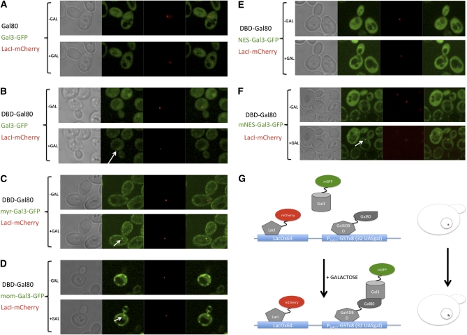
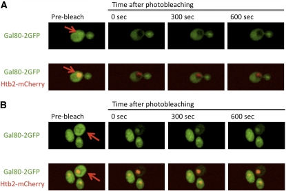
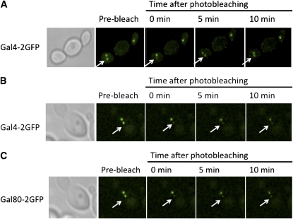
The yeast transcriptional activator Gal4 localizes to UAS(GAL) sites even in the absence of galactose but cannot activate transcription due to an association with the Gal80 protein. By 4 min after galactose addition, Gal4-activated gene transcription ensues. It is well established that this rapid induction arises through a galactose-triggered association between the Gal80 and Gal3 proteins that decreases the association of Gal80 and Gal4. How this happens mechanistically remains unclear. Strikingly different hypotheses prevail concerning the possible roles of nucleocytoplasmic distribution and trafficking of Gal3 and Gal80 and where in the cell the initial Gal3-Gal80 association occurs. Here we tested two conflicting hypotheses by evaluating the subcellular distribution and dynamics of Gal3 and Gal80 with reference to induction kinetics. We determined that the rates of nucleocytoplasmic trafficking for both Gal80 and Gal3 are slow relative to the rate of induction. We find that depletion of the nuclear pool of Gal3 slows the induction kinetics. Thus, nuclear Gal3 is critical for rapid induction. Fluorescence-recovery-after-photobleaching experiments provided data suggesting that the Gal80-Gal4 complex exhibits kinetic stability in the absence of galactose. Finally, we detect Gal3 at the UAS(GAL) only if Gal80 is covalently linked to the DNA-binding domain. Taken altogether, these new findings lead us to propose that a transient interaction of Gal3 with Gal4-associated Gal80 could explain the rapid response of this system. This notion could also explain earlier observations.


Figures

Figure 1
Figure 1
Gal3 distribution does not change in the course of early induction. (A) Sc859 (Gal3-2GFP) cells expressing Htb2-mCherry from pFJ35 were grown to mid-log phase in glycerol–lactic acid media and were placed into the Cellasik Onyx microfluidics system. Prior to imaging, cells were treated with cycloheximide for 15 min to prevent galactose-induced increases in Gal3-GFP. Images were acquired before and 45 min after galactose induction. (B) Sc723 cells expressing Hog1-GFP from pPS1739 and Htb2-mCherry from pFJ35 were grown to mid-log phase and were placed into the Cellasik Onyx microfluidics system. Images were acquired before and 10 min after osmotic stress with 500 mM NaCl.
Figure 2
Figure 2
Gal3 does not move into the nucleus rapidly. (A and B) Yeast strain Sc859 nuclei, defined by the nuclear marker Htb2-mCherry from pFJ35, were bleached briefly. The recovery of GFP signal from Gal3-GFP was monitored up to 10 min, at which point only 19% re-equilibration was observed. (C and D) RLY2800 (Fus3-GFP) cells were grown to mid-log phase in glycerol–lactic media. Distribution of Fus3-GFP was imaged, and the indicated nucleus was bleached. The nuclear:cytoplasmic ratio of the molecules completely re-equilibrated <20 sec after bleaching.
Figure 3
Figure 3
Effect of different tags on Gal3 distribution. Sc724 (gal3Δ) cells expressing (A) NES-Gal3-GFP from pOE183, (B) mNES-Gal3-GFP from pOE185, (C) myr-Gal3-GFP from pGP56GFP, or (D) mom-Gal3-GFP from pGP57GFP. All cells were grown to mid-log phase in glycerol–lactic media. (E) The ratio of the nuclear:cytoplasmic signal exhibited by the indicated molecule tagged with GFP was determined by measuring signal density. Ten cells were evaluated for each molecular species, and the results were averaged. The cells were induced for 4 hr in 2% galactose to enhance the GFP signal and were shifted back to gly/lac for 4 hr, at which time image acquisition began. The nuclear regions were marked by Htb2-mCherry that was expressed from pFJ35. (F) Total cellular levels of the various Gal3 molecules in the whole-cell extracts are demonstrated by the Western blot.
Figure 4
Figure 4
Effect of depleting nuclear Gal3 on induction kinetics. Sc724 (gal3Δ) cells were transformed with pOE33 (PGAL1-2GFP) and pAKS15 (Gal3WT), pOE180 (NES-Gal3), or pOE182 (mNES-Gal3). Cultures were grown to mid-log phase in glycerol–lactic acid media and induced for indicated times. The GFP produced at each time point was determined by a Western blot. A nonspecific band from the same blot served as a loading control.
Figure 5
Figure 5
Effect of mis-localization tags on the nuclear Gal3 pool. In Sc932 (gal3Δ, [PGAL1-GSTx8]::[LacOx64]::LEU2) cells, (A) Gal80 or (B–F) DBD-Gal80 was expressed from pOE142 or pOE165, respectively. The indicated Gal3 species were expressed from (A and B) pGP17, (C) pGP56GFP, (D) pGP65, (E) pOE183, or (F) pOE185. (G) Schematic presentation of the array system. The cells were grown in glycerol–lactic to mid-log phase and placed into chambers of Cellasik Microfluidics plates. Synthetic drop-out media with 3% glycerol and 2% lactic acid as carbon source were fed through the chambers for 20 min. Images of cells were obtained before and at 1 hr after galactose addition. The same cells were monitored for Gal3-GFP spots, which colocalized with LacI-mCherry expressed from pME9.
Figure 6
Figure 6
Subcellular distribution and dynamics of Gal80. Sc862 (Gal80-2GFP) cells expressing Htb2-mCherry from pFJ35 were grown to mid-log phase in glycerol–lactic acid media. The cells were induced with galactose for 4–6 hr to enhance the GFP signal and then shifted back to non-inducing media for 4 hr. The (A) nuclear or (B) cytoplasmic region was briefly bleached.
Figure 7
Figure 7
NLS-Gal80-GFP does not shuttle rapidly in heterokaryons. NLS-Gal80-GFP was expressed in wild-type MATa cells from a GAL1/CYC1 promoter. The expression was repressed with 2% glucose for 2 hr, after which the cells were mated with MATα kar1-1 cells. Binucleate heterokaryons appeared within 2 hr. The GFP signal was found in both nuclei of the heterokaryons. One of the two nuclei was bleached briefly, and the heterokaryon nuclei were monitored for 5 min.
Figure 8
Figure 8
Dynamics of Gal4–DNA and Gal80–Gal4 interactions. (A and B) Sc930 (MATa Gal4-2GFP [PGAL1-GSTx8]::[LacOx64]::LEU2) and (C) Sc920 (MATa Gal80-2GFP [PGAL1-GSTx8]::[LacOx64]::LEU2) strains were mated with Sc965 (MATα Gal4-2GFP [PGAL1-GSTx8]::[LacOx64]::LEU2) and Sc963 (MATα Gal80-2GFP [PGAL1-GSTx8]::[LacOx64]::LEU2), respectively. The resulting diploid cells containing two arrays were grown to mid-log phase in (A) galactose or (B and C) glycerol–lactic acid media. One of the GFP spots in the cells was briefly bleached, and the cells were monitored for 10 min. (A) The Gal4-2GFP signal in the bleached array of the cells grown with galactose recovered completely within 5 min, while no detectable re-equilibration was observed for (B) Gal4-2GFP and (C) Gal80-2GFP spots in cells grown in glycerol–lactic acid media.

References

    1. Ansari A. Z., Reece R. J., Ptashne M., 1998. A transcriptional activating region with two contrasting modes of protein interaction. Proc. Natl. Acad. Sci. USA 95: 13543–13548 - PMC - PubMed
    1. Arndt K. M., Ricuperohovasse S., Winston F., 1995. Tbp mutants defective in activated transcription in-vivo. EMBO J. 14: 1490–1497 - PMC - PubMed
    1. Bhaumik S. R., Green M. R., 2001. SAGA is an essential in vivo target of the yeast acidic activator Gal4p. Genes Dev. 15: 1935–1945 - PMC - PubMed
    1. Blackwell E., Halatek I. M., Kim H. J. N., Ellicott A. T., Obukhov A. A., et al. , 2003. Effect of the pheromone-responsive G(alpha) and phosphatase proteins of Saccharomyces cerevisiae on the subcellular localization of the Fus3 mitogen-activated protein kinase. Mol. Cell. Biol. 23: 1135–1150 - PMC - PubMed
    1. Blank T. E., Woods M. P., Lebo C. M., Xin P., Hopper J. E., 1997. Novel Gal3 proteins showing altered Gal80p binding cause constitutive transcription of Gal4p-activated genes in Saccharomyces cerevisiae. Mol. Cell. Biol. 17: 2566–2575 - PMC - PubMed

Publication types

MeSH terms

Substances