. 2011 Dec;39(22):9592-604.
doi: 10.1093/nar/gkr698. Epub 2011 Sep 2.

Signal-induced Brd4 release from chromatin is essential for its role transition from chromatin targeting to transcriptional regulation

Affiliations

Signal-induced Brd4 release from chromatin is essential for its role transition from chromatin targeting to transcriptional regulation

Nanping Ai et al. Nucleic Acids Res. 2011 Dec.

Abstract

Bromodomain-containing protein Brd4 is shown to persistently associate with chromosomes during mitosis for transmitting epigenetic memory across cell divisions. During interphase, Brd4 also plays a key role in regulating the transcription of signal-inducible genes by recruiting positive transcription elongation factor b (P-TEFb) to promoters. How the chromatin-bound Brd4 transits into a transcriptional regulation mode in response to stimulation, however, is largely unknown. Here, by analyzing the dynamics of Brd4 during ultraviolet or hexamethylene bisacetamide treatment, we show that the signal-induced release of chromatin-bound Brd4 is essential for its functional transition. In untreated cells, almost all Brd4 is observed in association with interphase chromatin. Upon treatment, Brd4 is released from chromatin, mostly due to signal-triggered deacetylation of nucleosomal histone H4 at acetylated-lysine 5/8 (H4K5ac/K8ac). Through selective association with the transcriptional active form of P-TEFb that has been liberated from the inactive multi-subunit complex in response to treatment, the released Brd4 mediates the recruitment of this active P-TEFb to promoter, which enhances transcription at the stage of elongation. Thus, through signal-induced release from chromatin and selective association with the active form of P-TEFb, the chromatin-bound Brd4 switches its role to mediate the recruitment of P-TEFb for regulating the transcriptional elongation of signal-inducible genes.


Figures

Figure 1.
Figure 1.
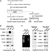
Almost all Brd4 is associated with chromatin in HeLa cells. (A) Schematics for modified nuclear fractionation (MONF) and DNase I digestion procedures. HeLa cells are suspended in buffer A, and the swollen cells are extracted three times with a low-salt buffer to yield low-salt fraction (LSF). The low-salt extracted nuclei (LSENs) are further extracted with a high-salt buffer to obtain high-salt fraction (HSF). Finally, the nuclear pellet is boiled in SDS-loading buffer, and saved as nuclear lysate fraction (NLF). Before further analysis, LSF, HSF and NLF are adjusted to the same volume and salt concentration. To verify that P-TEFb and its related factors in HSF reflected chromatin-bound components, LSENs are digested with DNase I and the supernatant is saved, whereas the nuclear pellet is subjected either to DNA isolation or to nuclear lysis. (B) The LSF, HSF and NLF fractions prepared from HeLa cells were analyzed by western blotting (WB) for the indicated proteins. (C) DNase I digestion assay of LSENs. Proteins in the supernatant and nuclear lysate after DNase I digestion of LSENs were analyzed by WB (right), and chromatin DNA was analyzed on ethidium bromide (EB) stained agarose gel (Left), with RNasin treatment as a control.
Figure 2.
Figure 2.
UV or HMBA treatment induces the release of Brd4 from chromatin. (A) LSF (left) and HSF (right) prepared from the UV- or HMBA-treated HeLa cells were subjected to western blotting (WB) for the indicated proteins. (B) Salt-titrated extraction assay of HeLa cells treated with HMBA or UV. The cells were extracted three times with low-salt buffer, and the nuclei were aliquoted and each was extracted with buffer containing indicated concentrations of NaCl. The nuclear pellets were lysed in SDS-loading buffer and analyzed by WB to determine the amount of Brd4 remained in chromatin. (C) The levels of released Brd4 in LSF (top) and the total amount of Brd4 in the cells (bottom) were determined by WB for HeLa cells that were treated with 10 mM HMBA for 4 h, and recovered in normal culture medium for the indicated period.
Figure 3.
Figure 3.
Both the chromatin-bound and signal-released Brd4 proteins associate with P-TEFb that contains phosphorylated CDK9 T-loop. (A) Levels of Brd4 and Cdk9 in anti-Flag immunoprecipitates from LSF (left) and HSF (right) of MCAP (HA-Brd4-f stable line) cells with indicated treatments were analyzed by western blotting (WB). (B) The levels of bulk and T186-phosphorylated (pT186) form of Cdk9 in anti-Flag immunoprecipitates derived from LSF (left) and HSF (right) of F1C2 (Cdk9-f stable line) cells treated with UV or HMBA were analyzed by WB. (C) Detection of Cdk9-T186 phosphorylation levels of Brd4-associated P-TEFb. Proteins were isolated from LSF and HSF of UV-treated MCAP cells by anti-Flag affinity purification. The levels of bulk and pT186 form of Cdk9 and indicated proteins were determined by WB, with HEXIM1-bound P-TEFb isolated from the LSF of HH8 (f-HEXIM1 stable line) cells as a control for the fully phosphorylated CDk9-T186. The sample volumes were adjusted to achieve equal loading of Cdk9 for the measurement of pT186 in (B, C).
Figure 4.
Figure 4.
Impeding Brd4 release from chromatin abolishes Brd4-mediated P-TEFb recruitment. (A) The effect of TSA treatment on stimulation-induced Brd4 release. Brd4 in LSFs prepared from HeLa cells treated with UV (left) or HMBA (right) either with or without TSA pretreatment as indicated were analyzed by western blotting (WB). (B) Salt-titrated extraction of HeLa cells treated with TSA and/or HMBA. The cells were extracted with low-salt buffer to remove chromatin-free Brd4, and subsequently the nuclei were extracted with buffer containing indicated concentrations of NaCl. The levels of released Brd4 were analyzed by WB. (C) Effect of TSA treatment on stimulation-induced nucleosomal histone H4 deacetylation. WB analysis of nucleosomal histone H4 acetylation levels (top) with quantification (bottom, averaged from three independent experiments) for HeLa cells treated with UV or HMBA either with or without TSA pretreatment as indicated. (D) Effect of TSA pretreatment on the levels of UV- or HMBA-induced Brd4/P-TEFb complex. Anti-Flag immunoprecipitates from LSF (left) and HSF (right) of MCAP (HA-Brd4-f stable line) cells with indicated treatments were analyzed by WB.
Figure 5.
Figure 5.
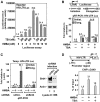
HDAC inhibitor blocks HMBA-stimulated HIV-1 transcriptional elongation. (A) Effect of HDAC inhibitor TSA on HMBA-stimulated HIV-1 expression. Luciferase activities of the cell lysates from HeLa cells with an integrated HIV-1 LTR-luciferase gene that were pretreated with the indicated amounts of TSA before incubated with HMBA were plotted based on three independent experiments. (B) The levels of transcripts corresponding to the regions of initiation (+1 to +59, TAR) or elongation (+496 to +593, luciferase) as illustrated on the top (45) were detected by qRT-PCR. HeLa cells with an integrated HIV-1 LTR-luciferase gene were pretreated with 400 nM TSA before incubated with 10 mM HMBA. The luciferase activities based on three independent experiments were plotted. (C) Effects of infection with lentivirus expressing scrambled shRNA or Brd4 shRNA on the levels of HMBA-induced HIV-1 transcription were measured by qRT-PCR (left) as in (B), and the results based on three independent experiments were plotted, with those untreated with HMBA setting to 1. The efficiency of Brd4 knockdown was analyzed by WB with β-actin as the loading control (right). (D) Chromatin immunoprecipitation (ChIP) assay with anti-Cdk9 antibody for HeLa cells with an integrated HIV-1 LTR-luciferase gene treated with TSA and/or HMBA. The precipitated DNA was analyzed by real-time PCR with the primers matching to the promoter region (top), and plotted as the percentage of input (bottom) based on two independent experiments.
Figure 6.
Figure 6.
The forced release of endogenous Brd4 from chromatin by ΔC-mutant of Brd4 augments HIV-1 transcription. (A) The levels of endogenous Brd4 in LSFs from HeLa cells transfected with empty vector, HA-tagged ΔC-mutant of Brd4 (HA-ΔC) or HA-tagged PID (P-TEFb-interacting domain) of Brd4 (HA-PID) were analyzed by western blotting (WB). The levels of HA-ΔC and HA-PID in cell lysates were also detected by WB (bottom). (B) Luciferase activities of HIV-LTR-Luc integrated HeLa cells transfected with indicated Brd4 mutant constructs were plotted based on three independent experiments.
Figure 7.
Figure 7.
A model depicting the signal-induced functional transition of Brd4 from chromatin targeting to transcriptional regulation. In the absence of the external stimuli, almost all Brd4 is associated with acetylated chromatin (indicated as acetyl-nucleosome). The signals trigger histone H4 deacetylation (red dash line) by activating yet-to-be identified HDAC(s) (red question mark), which causes the release of Brd4 from chromatin (black dash line). Meanwhile, the signals also induce the dephosphorylation of Cdk9 at T186 (blue dash line) by the cooperative actions of the activated phosphatases (PP2B and PP1α), thereby liberating P-TEFb from the inactive 7SK snRNP (19). Through selective association with the transcriptional active form of P-TEFb that has its Cdk9T-loop re-phosphorylated by unknown kinase(s) (blue question mark), the released Brd4 mediates the recruitment of active P-TEFb to promoter-proximal region (green dash line), where P-TEFb modulates the processivity of Pol II, thereby leading to productive elongation. This recruitment is most likely through recognizing adaptor factors, such Mediator or sequence-specific transcription factors.

Similar articles

Cited by

References

    1. Selth LA, Sigurdsson S, Svejstrup JQ. Transcript Elongation by RNA polymerase II. Annu. Rev. Biochem. 2010;79:271–293. - PubMed
    1. Peterlin BM. Transcription elongation takes central stage: the P-TEFb connection. Cell Cycle. 2010;9:2933–2934. - PubMed
    1. Price DH. P-TEFb, a cyclin-dependent kinase controlling elongation by RNA polymerase II. Mol. Cell. Biol. 2000;20:2629–2634. - PMC - PubMed
    1. Price DH. Poised polymerases: on your mark … get set … go! Mol. Cell. 2008;30:7–10. - PubMed
    1. Zhou Q, Yik JH. The Yin and Yang of P-TEFb regulation: implications for human immunodeficiency virus gene expression and global control of cell growth and differentiation. Microbiol. Mol. Biol. Rev. 2006;70:646–659. - PMC - PubMed

Publication types

MeSH terms