. 2011 Dec;39(22):9705-19.
doi: 10.1093/nar/gkr669. Epub 2011 Sep 2.

Activity, specificity and structure of I-Bth0305I: a representative of a new homing endonuclease family

Affiliations

Activity, specificity and structure of I-Bth0305I: a representative of a new homing endonuclease family

Gregory K Taylor et al. Nucleic Acids Res. 2011 Dec.

Abstract

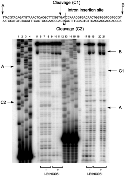
Novel family of putative homing endonuclease genes was recently discovered during analyses of metagenomic and genomic sequence data. One such protein is encoded within a group I intron that resides in the recA gene of the Bacillus thuringiensis 03058-36 bacteriophage. Named I-Bth0305I, the endonuclease cleaves a DNA target in the uninterrupted recA gene at a position immediately adjacent to the intron insertion site. The enzyme displays a multidomain, homodimeric architecture and footprints a DNA region of ~60 bp. Its highest specificity corresponds to a 14-bp pseudopalindromic sequence that is directly centered across the DNA cleavage site. Unlike many homing endonucleases, the specificity profile of the enzyme is evenly distributed across much of its target site, such that few single base pair substitutions cause a significant decrease in cleavage activity. A crystal structure of its C-terminal domain confirms a nuclease fold that is homologous to very short patch repair (Vsr) endonucleases. The domain architecture and DNA recognition profile displayed by I-Bth0305I, which is the prototype of a homing lineage that we term the 'EDxHD' family, are distinct from previously characterized homing endonucleases.


Figures

Figure 1.
Figure 1.
Features of the I-Bth0305I protein sequence. The catalytic domain of the protein is indicated in blue font with putative active site residues in red. The underlined region corresponds to the sequence logo in the lower blue frame, where the predicted active sites are marked by red bullets. Two repeats of ‘NUMOD’ sequence motifs in the putative DNA-binding domain are shown in pink underlined font, with their motif logo shown above in the upper pink frame. Logo positions are numbered according to I-Bth0305I. Gaps (–) mark deletions in I-Bth0305I relative to other protein family members, and I-Bth0305I residue Trp149 is an insert relative to the family members and thus not shown in the logo. Beneath the logos of the repeated NUMOD motif is its predicted structure (cylinders for α-helices and arcs for loops and turns). The hatched region denotes a predicted DNA-binding HTH motif.
Figure 2.
Figure 2.
Determination of the I-Bth0305I DNA target site. (A) I-Bth0305I cleaves a DNA target site containing the sequence of the RecA host gene spanning the intron insertion site. (B) In competition digests, three substrates (one corresponding to the uninterrupted allele of the bacteriophage RecA gene; ‘intron-minus’ and two substrates corresponding to the intron-containing allele of the same RecA gene; ‘intron-plus’) were simultaneously digested with 70 nM I-Bth0305I. Only the intron-minus allele of the RecA sequence is cleaved. (C) Sequencing of the most strongly nicked and cleaved products resulting from a digest of lambda phage DNA with I-Bth0305I results in a specificity profile (i.e. a ‘logo’ plot) indicating that the strongest features of substrate specificity correspond to the pseudopalindromic consensus sequence 5′-TTxG-x6-CxAA-3′, which is cleaved on each strand to give two base, 5′ overhangs centered in the middle of the symmetric DNA target. For the logo shown in this figure, only those sites in the lambda genome displaying 90% or higher cleavage of at least one strand were included in the creation of the consensus sequence. Reducing the cleavage threshhold for inclusion of more sequences in the determination of the enzyme's specificity profile quantitatively affects the absolute values for information content at individual positions, but does not alter the consensus sequence identity. (D) Sequence of the recA target region of the I-Bth0305I endonuclease that is cleaved by I-Bth0305I. The results of run-off sequencing of cleaved top and bottom strands is consistent with generation of two base, 5′ cohesive overhangs observed in the prior experiment with lambda genomic DNA. The target site is numbered to illustrate the two pseudosymmetric half-sites in the recA gene target that flank the middle of the cleavage site. Following convention for homing endonuclease target site numbering, the left half site is accorded with negative position numbering, and the right half-site is accorded with positive position numbering. Black bullets indicate positions that are palindromically conserved between the left and right half-sites.
Figure 3.
Figure 3.
I-Bth0305I target site DNAse I footprint. Forward and reverse PCR primers were labeled with 32P and used to generate PCR products labeled at either end. In lanes 6–8 and 17–18, reverse and forward labeled PCR products were digested with DNAse-I. In lanes 9–11 and 19–21, labeled PCR products were incubated with 20 µM I-Bth0305I and digested with DNAse I. Through a comparison of the I-Bth0305I protected and unprotected DNAse I ladders, a 60 base region with the site of catalysis at its center is protected from DNAse I degradation by specific binding of I-Bth0305I. Lanes 1–4 and 12–15 are sequence ladders and lanes 5 and 16 are undigested PCR product.
Figure 4.
Figure 4.
Effect of multiple base pair substitutions on DNA cleavage. (A) Enzymatic cleavage was assayed in complementary experiments where digests were performed using a DNA substrates containing the target site that were disrupted by either insertion of 2 bp at several positions, or by systematic transversion of three consecutive base pairs. For insertion disruptions, two adenosines were inserted at one of the positions indicated by the red arrows, thus generating substrates ‘a’ to ‘g’. In transversion disruptions, several sets of three consecutive nucleotides, each marked by a bracket, were inversed, thus generating substrates ‘A’ to ‘K’. (B) Cleavage products produced by digestion of substrates a–g. Product generation is significantly impaired for substrates b, c and d, corresponding to insertions of additional base pairs after positions −5, 0 and +2 in the RecA target site. (C) Cleavage products produced by digestion of substrates A–K. Product generation is significantly impaired for substrates F, G, H and I, corresponding to transversion of three consecutive base pairs in a region extending from position −5 to +6.
Figure 5.
Figure 5.
Effect of reduced length of RecA target site on DNA cleavage. (A) Four separate substrates, ranging in total length from 2200 to 1300 bp, that contained specific bacteriophage RecA target sequences of various lengths centered around the site of cleavage were digested with I-Bth0305I and cleavage was measured. The experiment was conducted twice, with the various RecA sequences embedded in DNA of different overall length, to ensure that measurable differences in cleavage were due solely to the length of the phage RecA sequence in the substrates. (B) Quantitation of cleavage product formation for each substrate in the presence of 70 nM I-Bth0305I.
Figure 6.
Figure 6.
Effect of single base pair substitutions on DNA cleavage. (A) Each bar represents the relative cleavability of a target site that is altered at 1 bp relative to the wild-type target. (B) Raw data for cleavage specificity at positions −5 and −1, respectively. In these experiments, increasing concentrations of enzyme are used in competition experiments against equimolar concentrations of four DNA substrates that differ in length and in the identity of a single base pair at one position in the target. For each position being tested, the effect of DNA base pair mismatches were measured multiple times, including experiments in which the length of the substrates was reversed relative to the identity of the variable base pair (to ensure that differences in cleavage are due only to the sequence of the target).
Figure 7.
Figure 7.
Structural analyses of I-Bth0305I nuclease domain. (A) The crystal structure of the I-Bth0305I catalytic domain (PDB ID 3r3p). (B) The structure of E. coli Vsr endonuclease in the absence of bound DNA (PDB ID 1vsr). The bound zinc ion in the Vsr structure is the cyan sphere. (C) Superposition of the I-Bth0305I nuclease domain and the unbound Vsr endonuclease core. (D) Superposition of Vsr endonuclease active site and the putative I-Bth0305I active site and catalytic residues. (E) Side-by-side comparison of the I-Bth0305I nuclease domain and the DNA-bound structure of the Vsr endonuclease (PDB ID 1cw0) in the same relative orientations. In the Vsr-DNA co-crystal structure, the T:G mismatched nucleotide bases are shown in orange; the tryptophan residues (W68 and W86) that intercalate next to those mismatched DNA bases are shown in light blue and the active site magnesium ions are green spheres.
Figure 8.
Figure 8.
Conservation of the RecA DNA cleavage and intron insertion site and the RecA protein sequence. (A) Cartoon of the proposed domain architecture and DNA contact pattern exhibited by I-Bth0305I. The sequence (60 bp in length) corresponds to the overall region of DNA protected by the bound enzyme in DNAse I digestion experiments. Red bases are those that are recognized most specifically by the enzyme. (B) A logo plot indicating conservation of 1368 recA genes as described in the text. (C) Corresponding logo plot of translated RecA protein sequences from the same collection of sequences. The sequence of the 0305ϕ8–36 bacteriophage recA gene and RecA protein are shown between the two logo plots, with the intron insertion site and HEG insertion and cleavage sites on each DNA strand indicated. The recA coding sequence shown in this figure corresponds to the 60-bp region protected by bound I-Bth0305I in the DNAseI footprint experiment (Figure 3). The purple bases are the central 14 bp that display the most significant sequence specificity in cleavage assays. Black bullets between sequences of the two strands indicate the bases with palindromic symmetry between left and right DNA half-sites (also shown in Figure 2). The protein residues in the bacteriophage protein that correspond to the most conserved residues in the RecA proteins logo are underlined. The RecA L2 DNA-binding motif is indicated beneath the logo in panel b. (D) Structure of the E. coli RecA protein, bound as a filament on a single-stranded DNA target (pdb ID 3CMU) (33). The ssDNA ligand is in grey, with the RecA filament in blue and L2 regions in red.

Similar articles

Cited by

References

    1. Stoddard BL. Homing endonucleases: from microbial genetic invaders to reagents for targeted DNA modification. Structure. 2011;19:7–15. - PMC - PubMed
    1. Stoddard B, Belfort M. Social networking between mobile introns and their host genes. Mol. Microbiol. 2011;78:1–4. - PMC - PubMed
    1. Chevalier B, Monnat RJ, Jr, Stoddard BL. In: Homing Endonucleases and Inteins. Belfort M, Wood D, Derbyshire V, Stoddard B, editors. Vol. 16. Berlin: Springer Verlag; 2005. pp. 34–47.
    1. Flick KE, Jurica MS, Monnat RJ, Jr, Stoddard BL. DNA binding and cleavage by the nuclear intron-encoded homing endonuclease I-PpoI. Nature. 1998;394:96–101. - PubMed
    1. Zhao L, Bonocora RP, Shub DA, Stoddard BL. The restriction fold turns to the dark side: a bacterial homing endonuclease with a PD-(D/E)-XK motif. EMBO J. 2007;26:2432–2442. - PMC - PubMed

Publication types

Associated data