. 2011 Dec;39(22):9768-78.
doi: 10.1093/nar/gkr664. Epub 2011 Sep 2.

Influence of ground-state structure and Mg2+ binding on folding kinetics of the guanine-sensing riboswitch aptamer domain

Affiliations

Influence of ground-state structure and Mg2+ binding on folding kinetics of the guanine-sensing riboswitch aptamer domain

Janina Buck et al. Nucleic Acids Res. 2011 Dec.

Abstract

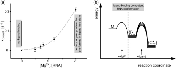
Riboswitch RNAs fold into complex tertiary structures upon binding to their cognate ligand. Ligand recognition is accomplished by key residues in the binding pocket. In addition, it often crucially depends on the stability of peripheral structural elements. The ligand-bound complex of the guanine-sensing riboswitch from Bacillus subtilis, for example, is stabilized by extensive interactions between apical loop regions of the aptamer domain. Previously, we have shown that destabilization of this tertiary loop-loop interaction abrogates ligand binding of the G37A/C61U-mutant aptamer domain (Gsw(loop)) in the absence of Mg(2+). However, if Mg(2+) is available, ligand-binding capability is restored by a population shift of the ground-state RNA ensemble toward RNA conformations with pre-formed loop-loop interactions. Here, we characterize the striking influence of long-range tertiary structure on RNA folding kinetics and on ligand-bound complex structure, both by X-ray crystallography and time-resolved NMR. The X-ray structure of the ligand-bound complex reveals that the global architecture is almost identical to the wild-type aptamer domain. The population of ligand-binding competent conformations in the ground-state ensemble of Gsw(loop) is tunable through variation of the Mg(2+) concentration. We quantitatively describe the influence of distinct Mg(2+) concentrations on ligand-induced folding trajectories both by equilibrium and time-resolved NMR spectroscopy at single-residue resolution.


Figures

Figure 1.
Figure 1.
Crystal structure of the G37A/C61U-mutant (Gswloop) from the B. subtilis guanine-sensing riboswitch aptamer domain in complex with the ligand thioguanine (thioG); (a) ribbon representation of the 3D structure of the Gswloop–thioG complex (chain a). The ligand thioG is presented in yellow dots; helices P1, P2 and P3 are color coded in blue, ligand-binding core region and loop regions are color coded in red; cobalt hexamine ions are shown in yellow stick representation; (b) Close-up view (rotated by 90° along vertical axis) of tertiary loop–loop interactions (RNA backbone of residues 32–38 in L2 and 60–66 in L3 are given; gray: chain a, blue: chain b). Residues of inter-helical base quadruples are shown in stick representation (arrows indicate 5′- to 3′-helix direction); (c) stick representation of the inter-helical base quadruple, including the mutations; black dashed lines represent hydrogen bonds, mutated inter-base pair angle is annotated in gray [U34(O2)–A65(N6)–U61(O2) ∼117°]; (d) close-up view of ligand-binding region from crystal structure of Gswloop–thioG. Local heterogeneity can be observed in molecules of one asymmetric unit in agreement with NMR spectra (Supplementary Figure S2) (gray: chain a, blue: chain b, with respective atom distances annotated).
Figure 2.
Figure 2.
Hypoxanthine- and Mg2+-induced effects on conformation and folding of Gswloop. (a) Mg2+ binding by the Gswloop–thioG complex (pdb: 3RKF). RNA residues of Gswloop (sequence position/structural element (77)/P1, (26, 30)/P2, (55, 56, 67)/P3), (34, 61, 38)/loop region, (47)/binding pocket that show NMR imino proton chemical shift changes upon Mg2+ titration Δδ(0–33eq)>15 Hz (12) are annotated in stick representation on the crystal structure of the G37A/C61U-mutant; cobalt hexamine ions are found in close proximity to the respective residues in the crystal structure (cobalt hexamine ions are highlighted in yellow); (b) Mg2+ binding by the Gswapt–hypoxanthine complex [pdb: 1U8D (10)]. RNA residues of Gswapt (sequence position/structural element (77)/P1, (26, 30, 31)/P2, (55, 56, 67, 72)/P3), (37, 38)/loop region, (47)/binding pocket that show NMR imino proton chemical shift changes upon Mg2+ titration Δδ(0–33eq)>15 Hz (28) are annotated in stick representation on the crystal structure of the wild-type Gswapt–hypoxanthine complex. Cobalt hexamine ions are found in close proximity to the respective residues in the crystal structure (cobalt hexamine ions are highlighted in orange); (c) kinetics of Mg2+- and hypoxanthine-induced RNA–ligand complex formation. Spectral changes recorded for imino proton signal U81 over time [s] are illustrated (*): time point of injection (t ∼ 0 s) of Mg2+ and hypoxanthine; time resolution/1D spectrum ∼8.6 s; black: 1D-spectra before injection, red: 1D-spectra following injection); (d) addition of hypoxanthine leads to small chemical shift changes of the imino proton signal of nucleotide U81 in helix P1 (Δδ∼8.2 Hz) (overlay of 1H,15N-HSQC spectral region of U81/Gswloop at an [RNA]:[Mg2+] ratio of ∼1:7 in presence (red) and absence of hypoxanthine (black)); (e) addition of Mg2+ leads to significant chemical shift changes of U81 (overlay of 1H,15N-HSQC spectral region at different [RNA]:[Mg2+] ratios: Δδ(0–7eq)∼24.6 Hz and Δδ(0–20eq)∼49.1 Hz).
Figure 3.
Figure 3.
Ligand-induced folding of Gswloop ([RNA]:[ligand]:[Mg2+] ∼1:1:8, 700 MHz, 283 K). Normalized integrals over time [s] of exemplary imino proton signals of (a) nucleotide U49 within the ligand-binding core region and of (b) nucleotide G38 forming part of the long-range inter-helical base pairing interactions (dark gray solid line: monoexponential fit). (c) Secondary structure of the G37A/C61U-mutant of the guanine-sensing riboswitch aptamer domain of the B. subtilis xpt-pbuX operon (for further construct details, see Supplementary Data).
Figure 4.
Figure 4.
Dependence of the equilibrium of free Gswloop and Gswloop–hypoxanthine complex on Mg2+ concentration. Signals originating from nucleotide U51 can be detected only in the ligand-bound form, while signals from nucleotide U17 can be observed in the free (U17free) and the RNA–ligand complex (U17complex), however, with different chemical shifts. The signal of nucleotide U69 shows differences neither in intensity nor significantly in chemical shift in the two conformations. (a) Normalized integrals relative to the integral U17total = U17free + U17complex for imino proton signal of U17free, U17complex and U51 as a function of the [Mg2+]:[RNA] ratio. (b) Overlay of 1D NMR spectra (15N-edited) of Gswloop (15N-uridine labeled) in the presence of equimolar concentrations of hypoxanthine and varying concentrations of Mg2+ (signal-to-noise is normalized relative to signal of nucleotide U69).
Figure 5.
Figure 5.
Mg2+ dependence of the ligand-induced Gswloop folding; (a) overall time constant koverall [s−1] of ligand-induced RNA folding process as a function of [Mg2+]:[RNA] ratio (the error is given from replica measurement at [Mg2+]:[RNA] ∼7:1; additional data at higher [Mg2+]:[RNA] ratios >20:1 could not be obtained due to the limited time-resolution of the NMR measurements; gray dashed line: sigmoidal fit; the choice of the fitting function was motivated by assuming that koverall [s−1] approaches saturation at an [RNA]:[Mg2+] ratio of 1:20); (b) free activation energies of the Mg2+- and/or ligand-induced conformational transitions are schematically depicted. In the free G37A/C61U-mutant (M, absence of Mg2+), secondary structure elements but neither the loop–loop interaction nor the ligand-binding region are pre-formed and Gswloop cannot bind ligand (28). Through variation of the [RNA]:[Mg2+] ratio, the ligand-binding capability can be restored ({I}mg) as well as formation of the tertiary loop–loop interaction at high Mg2+ concentrations ([RNA]:[Mg2+] >1:18). In the schematic diagram, this observation leads to a conformational RNA ensemble, whose dynamic and structural properties are Mg2+ dependent (signified by {}). The addition of Mg2+ leads to a rapid RNA conformational change. The derivation is based on the experimental finding that the Mg2+-induced conformational transition, monitored for Gswloop at an [RNA]:[Mg2+] ratio <1:18 (M→{I}mg), is faster than ∼8–10 s. The conformational transitions induced by Mg2+ are very fast and associated with energetic barriers that are small compared to the barriers associated with ligand binding. The kinetics of ligand binding ({I}mg→{C*}mg) strongly dependent on the [RNA]:[Mg2+] ratio. The variation in kinetic rate constants from the [Mg2+] concentration implies Mg2+-dependent differences of the free activation energy.

Similar articles

Cited by

References

    1. Brion P, Westhof E. Hierarchy and dynamics of RNA folding. Annu. Rev. Biophys. Biomol. Struct. 1997;26:113–137. - PubMed
    1. Draper DE, Grilley D, Soto AM. Ions and RNA folding. Annu. Rev. Biophys. Biomol. Struct. 2005;34:221–243. - PubMed
    1. Woodson SA. Compact intermediates in RNA folding. Annu. Rev. Biophys. 2010;39:61–77. - PMC - PubMed
    1. Thirumalai D, Hyeon C. RNA and protein folding: common themes and variations. Biochemistry. 2005;44:4957–4970. - PubMed
    1. Garst AD, Batey RT. A switch in time: detailing the life of a riboswitch. Biochim. Biophys. Acta. 2009;1789:584–591. - PMC - PubMed

Publication types