. 2011 Dec;39(22):9473-97.
doi: 10.1093/nar/gkr691. Epub 2011 Sep 3.

Evolution of the deaminase fold and multiple origins of eukaryotic editing and mutagenic nucleic acid deaminases from bacterial toxin systems

Affiliations

Evolution of the deaminase fold and multiple origins of eukaryotic editing and mutagenic nucleic acid deaminases from bacterial toxin systems

Lakshminarayan M Iyer et al. Nucleic Acids Res. 2011 Dec.

Abstract

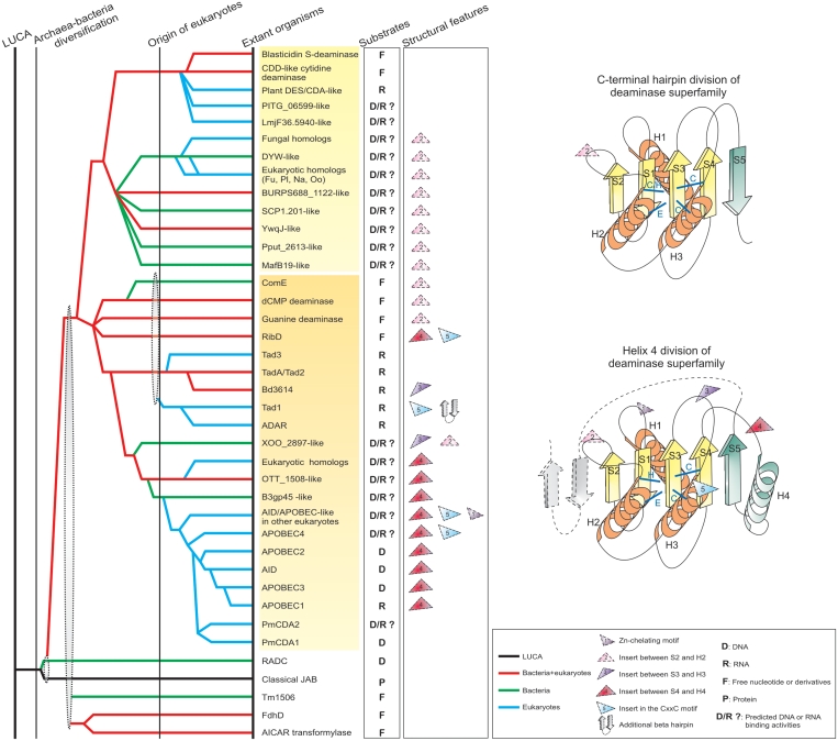
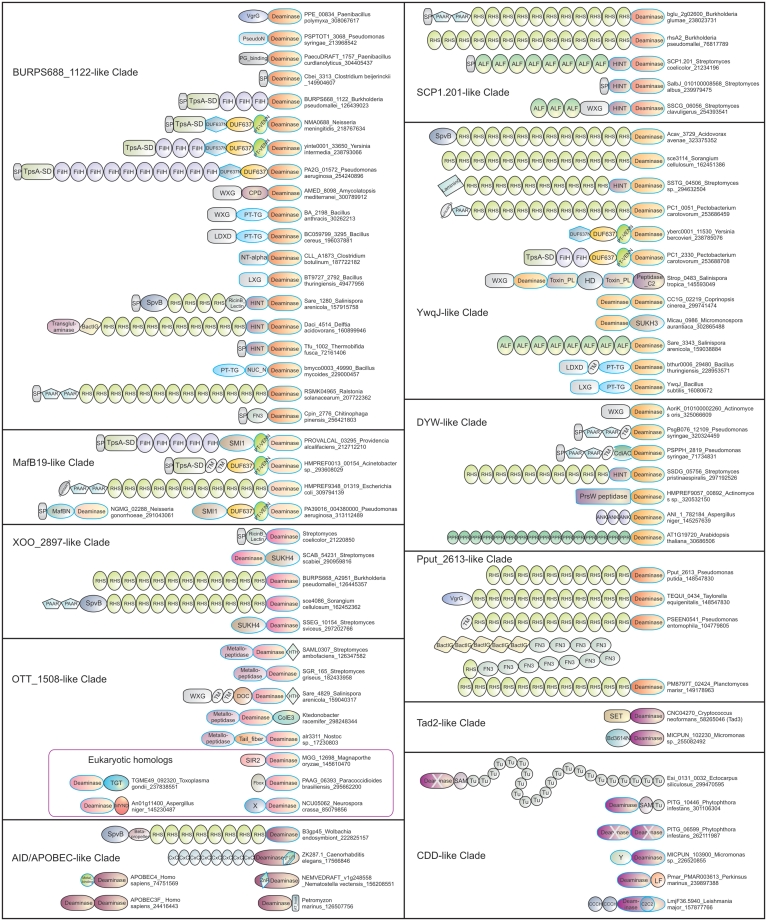
The deaminase-like fold includes, in addition to nucleic acid/nucleotide deaminases, several catalytic domains such as the JAB domain, and others involved in nucleotide and ADP-ribose metabolism. Using sensitive sequence and structural comparison methods, we develop a comprehensive natural classification of the deaminase-like fold and show that its ancestral version was likely to operate on nucleotides or nucleic acids. Consequently, we present evidence that a specific group of JAB domains are likely to possess a DNA repair function, distinct from the previously known deubiquitinating peptidase activity. We also identified numerous previously unknown clades of nucleic acid deaminases. Using inference based on contextual information, we suggest that most of these clades are toxin domains of two distinct classes of bacterial toxin systems, namely polymorphic toxins implicated in bacterial interstrain competition and those that target distantly related cells. Genome context information suggests that these toxins might be delivered via diverse secretory systems, such as Type V, Type VI, PVC and a novel PrsW-like intramembrane peptidase-dependent mechanism. We propose that certain deaminase toxins might be deployed by diverse extracellular and intracellular pathogens as also endosymbionts as effectors targeting nucleic acids of host cells. Our analysis suggests that these toxin deaminases have been acquired by eukaryotes on several independent occasions and recruited as organellar or nucleo-cytoplasmic RNA modifiers, operating on tRNAs, mRNAs and short non-coding RNAs, and also as mutators of hyper-variable genes, viruses and selfish elements. This scenario potentially explains the origin of mutagenic AID/APOBEC-like deaminases, including novel versions from Caenorhabditis, Nematostella and diverse algae and a large class of fast-evolving fungal deaminases. These observations greatly expand the distribution of possible unidentified mutagenic processes catalyzed by nucleic acid deaminases.


Figures

Figure 1.
Figure 1.
Representative structures of the deaminase fold. All structural cartoons are shown in an approximately similar orientation. The α-helices are colored purple, β-sheets yellow and loops gray. The predicted and known active site residues and substrates and ligands (if known) are labeled. The β-strand which adopts different orientations in the two major deaminase divisions is shown in dark green. Surface diagrams are colored based on their positions relative to the center of the structure (outside to inside: blue to red) to illustrate the binding cleft. For the JAB domain, only the relevant portion of the dimeric Ub-substrate that interacts with the active site is rendered. Similarly, for the AICAR transformylase only the region of the B chain (the other change of the dimeric unit) that interacts with the active site pocket is rendered.
Figure 2.
Figure 2.
Reconstructed evolutionary history for the deaminase fold and key structural features. On the left is a reconstructed evolutionary history of the deaminase fold. Individual lineages are listed to the right and grouped according to the classification given in the text and Table 1. The inferred evolutionary depth of the lineages is traced by solid horizontal lines across the relative temporal epochs representing major evolutionary transitional periods shown as vertical lines. Horizontal lines are colored according to their observed phyletic distributions; the key for this coloring scheme is given at the bottom right of the figure. Dashes indicate uncertainty in terms of the origins of a lineage, while gray ellipses group lineages of relatively restricted phyletic distribution with more broadly distributed lineages, indicating that the former likely underwent rapid divergence from the latter. Known and predicted functions of the deaminases are shown next to the clade names. On the right are topologies of the two major divisions within the deaminase superfamily. Insert positions characteristic of various deaminase lineages are marked in both the evolutionary history and topology diagrams. The β-strands and α-helices of the conserved deaminase core are colored yellow and orange respectively. Additional structural elements are colored dark green. Refer to the key for coloring schemes and abbreviations. Additionally, Fu: fungi, Pl: Plants, Na: Naegleria, Oo: Oomycetes.
Figure 3.
Figure 3.
Multiple alignment of the deaminase superfamily. Proteins are denoted by their gene name, species abbreviations and GI (Genbank Index) numbers separated by underscores and are further grouped by their familial associations, shown to the right of the alignment. Secondary structure assignments are shown above the alignment, where the green arrow represents the β-strand and the orange cylinder the α-helix. Helices not part of the universally conserved deaminase core are shown in a different color. Secondary structure was derived from a combination of crystal structures and alignment-based predictions. Inserts are replaced by the corresponding number of residues. Columns in the alignment are colored based on their amino acid conservation at 65% consensus. Residues shared by members of the AID/APOBEC clade are marked in a red box. A temporary id was assigned for the Emiliania huxleyi sequence and its complete sequence is available in the Supplementary Data. Red asterisks are placed at the end of sequences that are truncated and lack terminal secondary structure elements of the conserved deaminase core. The coloring scheme and consensus abbreviations are as follows: h, hydrophobic (ACFILMVWY); l, aliphatic (LIV) and a, aromatic (FWY) residues shaded yellow; b, big residues (LIYERFQKMW), shaded gray; s, small residues (AGSVCDN) and u, tiny residues (GAS), shaded green; p, polar residues (STEDKRNQHC) shaded blue; c, charged residues (DEHKR) shaded magenta and zinc coordinating residues shaded red. Strand-5 of the two distinct deaminase divisions are aligned separately given their independent emergence. Species abbreviations are as follows; Aae: Aquifex aeolicus; Acel: Acetivibrio cellulolyticus; Asp.: Actinomyces sp.; Ater: Aspergillus terreus; Atha: Arabidopsis thaliana; BPT4: Enterobacteria phage T4; Bbac: Bdellovibrio bacteriovorus; Bcer: Bacillus cereus; Bdor: Bacteroides dorei; Bpse: Burkholderia pseudomallei; Bsub: Bacillus subtilis; CAmo: Candidatus Amoebophilus; CKor: Candidatus Koribacter; Ccin: Coprinopsis cinerea; Cele: Caenorhabditis elegans; Cneo: Cryptococcus neoformans; Daci: Delftia acidovorans; Ecol: Escherichia coli; Ehux: Emiliania huxleyi; Hsap: Homo sapiens; Lmaj: Leishmania major; Lmon: Listeria monocytogenes; Mkan: Methanopyrus kandleri; Mory: Magnaporthe oryzae; Msp.: Micromonas sp.; Ncra: Neurospora crassa; Ngru: Naegleria gruberi; Nmen: Neisseria meningitidis; Nmul: Nakamurella multipartita; Nvec: Nematostella vectensis; Otsu: Orientia tsutsugamushi; Paer: Pseudomonas aeruginosa; Pbra: Pseudomonas brassicacearum; Pemar: Perkinsus marinus; Petma: Petromyzon marinus; Pinf: Phytophthora infestans; Plum: Photorhabdus luminescens; Pmar: Planctomyces maris; Pput: Pseudomonas putida; Psta: Pirellula staleyi; Psyr: Pseudomonas syringae; Rbel: Rickettsia bellii; Sare: Salinispora arenicola; Scel: Sorangium cellulosum; Scer: Saccharomyces cerevisiae; Scoe: Streptomyces coelicolor; Smoe: Selaginella moellendorffii; Tadh: Trichoplax adhaerens; Tequ: Taylorella equigenitalis; Tgon: Toxoplasma gondii; Tspi: Trichinella spiralis; Wend: Wolbachia endosymbiont; Xcam: Xanthomonas campestris; Xory: Xanthomonas oryzae.
Figure 4.
Figure 4.
Representative domain architectures of the deaminase superfamily. Proteins are denoted by their name, species and gi. Architectures are grouped based on the deaminase lineage in which they are present. Domains newly identified in this study are indicated by blue margin. For most part, standard domain names were used (as in PFAM). The various families of the SUKH superfamily of anti-toxins (e.g. Smi1, SUKH3 or SUKH4) are individually labeled. Other domain abbreviations: BactIG—a family of immunoglobulin fold domains found in bacteria; Bd3614N- N-terminal domain found in Bd3614-like deaminases; CPD—a Clostridium difficile Toxin A CPD type thiol peptidase; NT-α—N-terminal α-helical domain limited to firmicutes; PG_binding: peptidoglycan binding; various PT domains are pre-toxin domains; PseudoN—N-terminal domain limited to Pseudomonas; TM—transmembrane; Toxin_PL—Predicted papain-like peptidase toxin; SP—signal peptide; Tail_Fiber, a phage tail fiber-like peptidase; Tu—tudor; X—uncharacterized globular domains; Y—novel Rossmann fold domain. MafBN is a Neisseria-specific domain involved in toxin delivery along with the MafA lipoprotein.
Figure 5.
Figure 5.
Gene neighborhoods and contextual connection network of the deaminase superfamily. (A) Individual genes are represented as arrows pointing from the 5′- to the 3′-end of the coding frame. Genes were named according to their domain architectures. For each operon, the gene name, species name and gi of the deaminase (marked with a star) are indicated. Uncharacterized genes are shown as small gray boxes. Where possible, secretion pathways are indicated. Smi1, SUKH3 and SUKH4 are different clades of immunity proteins belonging to the SUKH superfamily. (B) Domains linked in a polypeptide are indicated by solid lines, whereas, contextual linkages between genes in operons are indicated by dashes of different colors. Lines are colored based on the deaminase clade. Black arrows indicate the polarity of domain arrangement in a polypeptide with the arrowhead pointing to the C-terminus, and white arrows show the order of genes in operons from 5′ to 3′. Multiple copies of domains or their direct linkages in operon are shown with arrow cycles. Key protein domains that correspond to diverse secretion systems (T5SS, T2SS, T7SS, T6SS, PVC, PrsW and the terminase system) are grouped together. Different deaminase clades are labeled with deaminase followed by numbers from 1 to 12. Toxin domains that are present in polytoxins are linked with bold lines. For domain abbreviations, please refer to Figure 4 legend.

References

    1. Harold CS. RNA and DNA Editing: Molecular Mechanisms and Their Integration into Biological Systems. Hoboken, New Jersey: John Wiley & Sons, Inc; 2008.
    1. Weiss B. The deoxycytidine pathway for thymidylate synthesis in Escherichia coli. J. Bacteriol. 2007;189:7922–7926. - PMC - PubMed
    1. Kumasaka T, Yamamoto M, Furuichi M, Nakasako M, Teh AH, Kimura M, Yamaguchi I, Ueki T. Crystal structures of blasticidin S deaminase (BSD): implications for dynamic properties of catalytic zinc. J. Biol. Chem. 2007;282:37103–37111. - PubMed
    1. Stenmark P, Moche M, Gurmu D, Nordlund P. The crystal structure of the bifunctional deaminase/reductase RibD of the riboflavin biosynthetic pathway in Escherichia coli: implications for the reductive mechanism. J. Mol. Biol. 2007;373:48–64. - PubMed
    1. Hamilton CE, Papavasiliou FN, Rosenberg BR. Diverse functions for DNA and RNA editing in the immune system. RNA Biol. 2010;7:220–228. - PubMed

Publication types