. 2011 Sep 2;30(21):4398-413.
doi: 10.1038/emboj.2011.315.

Orientation-specific signalling by thrombopoietin receptor dimers

Affiliations

Orientation-specific signalling by thrombopoietin receptor dimers

Judith Staerk et al. EMBO J. .

Abstract

Ligand binding to the thrombopoietin receptor is thought to stabilize an active receptor dimer that regulates megakaryocyte differentiation and platelet formation, as well as haematopoietic stem cell renewal. By fusing a dimeric coiled coil in all seven possible orientations to the thrombopoietin receptor transmembrane (TM)-cytoplasmic domains, we show that specific biological effects and in vivo phenotypes are imparted by distinct dimeric orientations, which can be visualized by cysteine mutagenesis and crosslinking. Using functional assays and computational searches, we identify one orientation that represents the inactive dimeric state and another similar to a physiologically activated receptor. Several other dimeric orientations are identified that induce proliferation and in vivo myeloproliferative and myelodysplastic disorders, indicating the receptor can signal from several dimeric interfaces. The set of dimeric thrombopoietin receptors with different TM orientations may offer new insights into the activation of distinct signalling pathways by a single receptor and suggests that subtle differences in cytokine receptor dimerization provide a new layer of signalling regulation that is relevant for disease.


Conflict of interest statement

The authors declare that they have no conflict of interest.

Figures

Figure 1
Figure 1
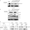
Design, validation and expression of seven fusion proteins that contain the Put3 coiled coil fused in all possible orientations to the TpoR TM–cytoplasmic domains. (A) Design of the cc-TpoR fusion proteins. The coiled-coil segment of Put3 was fused to the TM and cytoplasmic domains of TpoR. (B) By varying the junction between the Put3 coiled coil and the downstream TpoR TM α-helical domain, all seven possible registers could be imposed. All fusion proteins contain an HA tag at their N-terminus. The position of heptad repeats characteristic of coiled coils is denoted ag or a′–g′. The last residue of the Put3 coiled-coil is in a b position, while the first fused residue will be in a c position. The dimeric interface contains the a, d, d′ and a′ positions. (C) Crosslinking studies on live cells. Left panel: BOSC cells were transiently transfected with cDNA coding for seven truncated coiled-coil TpoR cysteine mutants (L505C). Cells were lysed after crosslinking with N,N′-1,2-phenylenedimaleimide (+ o-PDM), or in the absence of crosslinking (− o-PDM). Lysates were analysed on 12–14% Bis-Tris Gel followed by immunoblotting with an anti-HA antibody. Right panel: Helical representation of the different cc-TpoR interfaces and the putative localization of the Cys505 residue. (D) HA staining for cell surface expression of the cc-TpoR fusion proteins. The wtTpoR cell surface expression was set as 100%. Percent expression of the cc-TpoR fusion proteins was related to that of the wtTpoR. Shown is one experiment with data collected for 10 000 cells of three independent determinations; variations were <10%.
Figure 2
Figure 2
Cell proliferation induced by the seven cc-TpoR fusion proteins. (A, B) Proliferation assays were performed with Ba/F3 cells expressing the wtTpoR or the indicated cc-TpoR-0–VI fusion proteins (A), or K86P (KtoP) mutants thereof (B) at equal GFP levels in the absence of any cytokines or stimulated with 5 ng/ml Tpo as indicated. Cell numbers (averages of triplicates ±s.d.) were counted at day 6. One representative experiment out of four is shown. (C) Morphology of Ba/F3 cells expressing cc-TpoR-IV or cc-TpoR-I. Only cc-TpoR-IV induced cell-to-cell adhesion that lead to tight clusters (upper panel). Ba/F3 cells expressing the wtTpoR were stimulated with 5 or 500 ng/ml Tpo. At very high Tpo concentrations, cells formed clusters similar to those induced by cc-TpoR-IV (lower panel). Scale bar, 50 μm. (D) Proliferation assays were performed with UT7 cells expressing the wtTpoR or the cc-TpoR-0–VI fusion proteins at equal infection efficiency (GFP levels) in the absence of any cytokines or stimulated with 5 ng/ml Tpo, as indicated. Cell numbers were counted at day 8 and shown are averages of triplicates ±s.d. (E) Induction of CD41 expression of UT7 retrovirally transduced with the seven cc-TpoR fusion proteins or with the wtTpoR. Initiation of megakaryocyte differentiation of UT7 cells was assayed by flow cytometry analysis of the induction of cell surface CD41. The induction of CD41 cell surface localization by 5 ng/ml Tpo on cells expressing the wtTpoR was taken as 100%.
Figure 3
Figure 3
Transcriptional activity mediated by the wild-type (wt) TpoR or cc-TpoR-0–VI with and without alanine insertions. (A) Ba/F3 cells were selected in Tpo or without cytokines and starved overnight in RPMI/1 mg/ml BSA. To measure the transcriptional activity of STAT5 (left panel), STAT3 (middle) or MAP-kinase (MAPK) (right panel) pLHRE-luc, pGL3bPpr2-luc and pSRE-luc were used, respectively. pRL-TK encoding the renilla luciferase was co-electroporated for normalization of luciferase values. Cells were stimulated with 50 ng/ml Tpo or mock treated, as indicated. The results shown reflect averages of triplicate values ±s.d. Similar results were obtained performing three independent experiments. (B) γ2A cells were transfected with cDNAs coding for the cc-TpoR fusion proteins, JAK2 (left panel) or TYK2 (right panel), STAT5/STAT3 pGRR5-luc luciferase reporter and pRL-TK. The results represent averages of triplicate values ±s.d. of one representative experiment out of at least three. (C, D) γ2A cells were co-transfected with pGRR5-luc and pRL-TK luciferase reporter along with cDNAs coding for cc-TpoRs in which alanines were inserted at the end of the TM region, as indicated, and for JAK2 (C) or TYK2 (D).
Figure 4
Figure 4
Early signalling and traffic of cc-TpoR fusion proteins. (A) Immunoprecipitation of the indicated fusion proteins. Induction of phosphorylation of cytosolic Y626 was assessed using an anti-pY626 TpoR antibody. The anti-HA antibody was used to analyse the expression level of the cc-TpoR fusion protein. (B) Western blot analysis performed on Ba/F3 cells expressing the fusion proteins by using the indicated antibodies. (C) Recycling assay on Ba/F3 cells expressing cc-TpoR-I, cc-TpoR-II, cc-TpoR-III or cc-TpoR-IV. Cells were subjected to cell surface biotinylation, allowed to internalize the receptor, and then were maintained in culture in presence of JAK inhibitor I, in order to block JAK2 signalling, for the indicated time. Cell surface immunoprecipitation was performed on the cells followed by lysis in NP-40 buffer. Immunoprecipitates were analysed by immunoblot with an anti-biotin/HRP and an anti-HA.
Figure 5
Figure 5
In vivo expression of cc-TpoR fusion proteins. (A, B) Mice were reconstituted with bone marrow cells retrovirally transduced with the indicated cc-TpoR fusion proteins. Peripheral blood counts were measured 6–8 weeks after reconstitution. (A) Platelets counts are the average +s.d. of six mice per construct. (B) The peripheral number of platelets is shown for the indicated cc-TpoR fusion proteins; n, number of mice. Three independent reconstitutions are shown for TpoR wt, cc-TpoR-I and cc-TpoR-V, and two independent reconstitutions are shown for cc-TpoR-III and cc-TpoR-IV. (C) Peripheral blood was isolated from GFP-positive mice 6 weeks after reconstitution and blood smears were performed. Cover slides were stained using a May-Grunwald stain and analysed for qualitative or quantitative abnormalities in the blood. Bone marrow cytologic examination was performed after May Grunwald staining. The magnification was × 50. Scale bar, 50 μm.
Figure 6
Figure 6
Deletion of the juxtamembrane KWQFP motif eliminates differences in biologic effects between the seven cc-TpoR dimers. (A) Design of the cc-TpoR and Δ5cc-TpoR fusion proteins. The coiled-coil segment of Put3 was fused to the TM and cytoplasmic (CP) domains of the wtTpoR or the Δ5TpoR. (B, C) Proliferation assays were performed with Ba/F3 (A) and UT7 (B) cells expressing the cc-TpoR fusion proteins where the KWQFP motif was deleted (Δ5cc-TpoR-0–VI). Cell numbers (averages of triplicates ±s.d.) were counted at day 6 (A, B). (D) Cell morphology of Ba/F3 cells expressing Δ5cc-TpoR-IV or Δ5cc-TpoR-I. No clusters were induced by Δ5cc-TpoR-IV, which lacks the KWQFP motif. For all pictures shown, Ba/F3 cells were plated in 24-well plates and pictures were taken at day 4. (E) γ2A cells were transfected with cDNAs coding for the Δ5cc-TpoR fusion proteins, and TYK2, STAT3 and STAT5/STAT3 pGRR5-luc luciferase reporter and pRL-TK. The results represent averages of triplicate values ±s.d. of one representative experiment out of at least three. (F) Peripheral blood and bone marrow were isolated from mice expressing the indicated Δ5cc-TpoR fusion proteins. Coverslides were stained using a May-Grunwald stain and analysed for qualitative or quantitative abnormalities in the blood. Bone marrow was examined by haematoxylin–eosin staining. The magnification was × 50. Scale bar, 50 μm.
Figure 7
Figure 7
Computational searches of the TpoR transmembrane dimer. (A) Helical wheel predictions of the seven dimeric interfaces of the TM domain of the TpoR imposed by the Put3 coiled-coil fusion. Residue S498 of the TM domain is placed in the interface in cc-TpoR-I and cc-TpoR-V. (B) Computational search using the TpoR TM sequence from I485 to L506. The axial separation between the TM helices was set to be 10 Å. Computational searches with axial separations of 9.5–10.5 Å gave similar results. (C, D) Average molecular structures of the helix dimer corresponding to cc-TpoR-II and cc-TpoR-V. The helices have left-handed crossing angles and the dimer is viewed down the helix axis from the C-terminal end of the dimer. In cc-TpoR-II (C), the hydrogen bonding of Q509 stabilizes the helix dimer. The side chain of K509 contacts the aromatic ring of W508 characteristic of a stabilizing cation-π interaction. In cc-TpoR-V (D), the TM helices have rotated 50° counterclockwise, and hydrogen bonds form between indole NH of W508 and the amide side chain of Q509. The active cc-TpoR-I fusion protein adopts a conformation rotated by another 50° from cc-TpoR-V, where W508 residues would be predicted to clash.

References

    1. Abkowitz JL, Chen J (2007) Studies of c-Mpl function distinguish the replication of hematopoietic stem cells from the expansion of differentiating clones. Blood 109: 5186–5190 - PMC - PubMed
    1. Adams PD, Engelman DM, Brunger AT (1996) Improved prediction for the structure of the dimeric transmembrane domain of glycophorin A obtained through global searching. Proteins 26: 257–261 - PubMed
    1. Alexander WS, Maurer AB, Novak U, Harrison-Smith M (1996a) Tyrosine-599 of the c-Mpl receptor is required for Shc phosphorylation and the induction of cellular differentiation. EMBO J 15: 6531–6540 - PMC - PubMed
    1. Alexander WS, Metcalf D, Dunn AR (1995) Point mutations within a dimer interface homology domain of c-Mpl induce constitutive receptor activity and tumorigenicity. EMBO J 14: 5569–5578 - PMC - PubMed
    1. Alexander WS, Roberts AW, Nicola NA, Li R, Metcalf D (1996b) Deficiencies in progenitor cells of multiple hematopoietic lineages and defective megakaryocytopoiesis in mice lacking the thrombopoietic receptor c-Mpl. Blood 87: 2162–2170 - PubMed

Publication types

MeSH terms

Substances