. 2011 Sep 4;12(10):1010-6.
doi: 10.1038/ni.2095.

The inflammasome adaptor ASC regulates the function of adaptive immune cells by controlling Dock2-mediated Rac activation and actin polymerization

Affiliations

The inflammasome adaptor ASC regulates the function of adaptive immune cells by controlling Dock2-mediated Rac activation and actin polymerization

Sirish K Ippagunta et al. Nat Immunol. .

Abstract

The adaptor ASC contributes to innate immunity through the assembly of inflammasome complexes that activate the cysteine protease caspase-1. Here we demonstrate that ASC has an inflammasome-independent, cell-intrinsic role in cells of the adaptive immune response. ASC-deficient mice showed defective antigen presentation by dendritic cells (DCs) and lymphocyte migration due to impaired actin polymerization mediated by the small GTPase Rac. Genome-wide analysis showed that ASC, but not the cytoplasmic receptor NLRP3 or caspase-1, controlled the mRNA stability and expression of Dock2, a guanine nucleotide-exchange factor that mediates Rac-dependent signaling in cells of the immune response. Dock2-deficient DCs showed defective antigen uptake similar to that of ASC-deficient cells. Ectopic expression of Dock2 in ASC-deficient cells restored Rac-mediated actin polymerization, antigen uptake and chemotaxis. Thus, ASC shapes adaptive immunity independently of inflammasomes by modulating Dock2-dependent Rac activation and actin polymerization in DCs and lymphocytes.


Figures
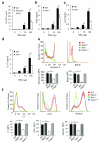
Figure 1
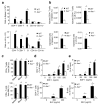
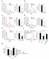
Figure 1. ASC controls antigen uptake and presentation independently of inflammasomes
a-d, Dendritic cells from naïve wild-type (WT) and Asc−/− mice (n = 4–6 mice per group) were co-cultured with WT CD4+ T cells for 72 hours in the presence of the indicated concentrations of BSA. Dose-dependent antigen-specific proliferation of lymphocytes was assessed by measuring uptake of [3H]-thymidine (a), and by analyzing the levels of the cytokines IFNγ (b), IL-6 (c) and IL-17 (d). *P < 0.01 (two-tailed Student’s t-test). Data are a representative of three independent experiments (mean ± s.d in a–d) (e) WT, Nlrp3−/−, Asc−/− and Casp1−/− bone marrow-derived dendritic cells (BMDCs) were incubated for 3 h at 37° C with fluorescein-labeled zymosan A or polystyrene beads. Phagocytosis was analyzed by flow cytometry. Results represent mean fluorescence intensity (MFI) ± S.E. of triplicates of at least three independent experiments. *P < 0.0005. (f) WT, Nlrp3−/−, Asc−/− and Casp1−/− BMDCs were incubated for 3 h at 37° C with fluorescein-labeled ovalbumin (OVA), dextran or luciferase yellow (LY). Macropinocytosis was anlayzed by flow cytometry. Results represent mean fluorescence intensity (MFI) ± S.E. of triplicates of at least three independent experiments. *P < 0.005.
Figure 2
Figure 2. ASC is required for lymphocyte migration in vitro and in vivo
(a) Spleen and lymph node cells of WT and Asc−/− mice were collected and stained for CD4, CD8, CD19, CD11b and CD11c. Data are expressed as the percentage of the total lymphocyte population in the spleen (upper panel) and axillary lymph nodes (lower panel) belonging to each category in WT (filled bars) and Asc−/− mice (open bars). (b) Naïve WT mice were injected with an equal ratio of congenically marked CD4+ T cells or B cells from WT and Asc−/− mice. 48 h after later, the spleen and axillary lymph nodes were collected to determine the levels of congenically marked WT and Asc−/− CD4+ T and B cells that migrated into these tissues. Results are expressed as the percentage of injected cells detected in the spleen or lymph nodes. (c) Mice were lethally irradiated and injected with an equal ratio of congenically marked WT and Asc−/− bone marrow. Blood, spleen and axillary lymph nodes were analyzed 6 weeks later for congenically marked CD4+ T cells and B cells derived from WT or Asc−/− bone marrow. Results are expressed as the percentage of the total bone marrow-derived lymphocytes. (d), (e) Migration of splenocytes of wild-type (WT) and Asc−/− mice was analyzed in vitro using a transwell chemotaxis assay. Data are expressed as the percentage of the total CD4+ T lymphocyte (d) and B cell (e) population migrating across the transwell. *P < 0.005. (n = 3–4 mice per group). Results represent means ± S.E. of triplicates of three independent experiments (a–e).
Figure 3
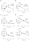
Figure 3. ASC is essential for antigen- and chemokine-induced Rac activation and actin polymerization in dendritic cells and lymphocytes, respectively
(a), (b) wild-type (WT) and Asc−/− BMDCs were treated with OVA for the indicated durations before Rac GTPase activity (a) and actin polymerization (b) were determined by flow cytometry. Results represent mean fluorescence intensity (MFI) ± S.E. of triplicates of at least three independent experiments. Baseline levels were arbitrarily assigned a value = 100. (c) CD4+ T cells and B cells isolated from spleens of WT and Asc−/− mice were treated in vitro with SDF-1 (500ng/mL) for the indicated durations before lysates were prepared and analyzed for Rac activation using a Rac1 G-LISA activation assay. (d) Isolated T and B cells from WT and Asc−/− mice were treated in vitro with SDF-1 (500ng/mL) for the indicated durations before actin polymerization was analyzed by flow cytometry. P-values <0.05 were considered significant (two-tailed Student’s t-test) (n = 1–3 per group) (a–d) Results represent means ± s.d. of triplicates of at least three independent experiments.
Figure 4
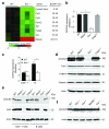
Figure 4. ASC regulates DOCK2 expression independently of inflammasomes and TLRs
(a) RNA from naïve wild-type (WT) and Asc−/− BMDCs was analyzed for gene expression using a HT MG-430 PM array plate. All genes that of which the transcript levels in WT and Asc−/− BMDCs differed three-fold or more are listed. (b) The mRNA expression levels of Dock2 in naïve WT, Nlrp3−/−, Asc−/− and caspase-1−/− BMDCs were quantified by qPCR and normalized against GAPDH. (c) DOCK2 expression levels in purified CD4+ T cells and B cells from WT and Asc−/− mice were determined by qPCR and normalized against GAPDH. (d) Western blot analysis of DOCK2, Fstl1, Fabp4, Galanin and Actin expression in naïve WT, Nlrp3−/−, Asc−/− and caspase-1−/− BMDCs. (e) CD4+ T cells and B cells from WT, Nlrp3−/−, Asc−/− and caspase-1−/− mice were purified and analyzed for protein expression of DOCK2 and ASC. (f) Lysates of naïve WT, Tlr2−/−, Tlr4−/−, MyD88−/− and Trif−/− BMDCs were analyzed for DOCK2 protein levels by Western blotting. Actin served as a loading control. Results are a representative of at least three independent experiments.
Figure 5
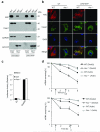
Figure 5. ASC localizes to the nucleus and controls Dock2 mRNA stability
(a) Wild-type (WT) and Asc−/− bone marrow-derived dendritic cells (BMDCs) were left untreated or primed with LPS (1 μg/mL) for 4 h, of which the final 15 minutes in the presence of ATP (5 mM). The cytosolic (Cyto) and nuclear (Nuc) compartments were separated and normalized for protein concentrations. Lysates were probed for expression of ASC and caspase-1 by Western blotting. GAPDH and Lamin B served as compartment-specific markers for the cytosolic and nuclear compartments, respectively. (b) WT and Asc−/− BMDCs were left untreated or primed with LPS (1 μg/mL) for 4 h, of which the final 15 minutes in the presence of ATP (5 mM). Cells were subsequently washed in PBS, fixed, permeabilized and stained for ASC and caspase-1 as described in Materials and Methods. DAPI was used to stain the nucleus. (c) WT and Asc−/− BMDCs were nucleofected with the pGL3 empty reporter vector (EV) or a pGL3 plasmid containing the Dock2 promoter (Dock2P). Luciferase activity was measured by scintillation counting. (d) WT and Asc−/− BMDCs were treated with 50μM DRB or 5μg/ml actinomycin-D before total RNA was prepared and expression levels of Dock2 and β-actin mRNA were determined by RT-qPCR and normalized against GAPDH. Baseline levels were arbitrarily assigned a value = 100. The t1/2 of mRNA stability was calculated as the time required for decay of 50% of baseline mRNA levels. *P < 0.005 (Student’s t-test) (d). Data presented represent means ± s.d. from one out of three (d) or four (e) independent experiments.
Figure 6
Figure 6. DOCK2 is critical for antigen uptake by dendritic cells, and restores immune cell functions in the absence of ASC
(ac) WT and Dock2−/− bone marrow-derived dendritic cells (BMDCs) were incubated for 3 h at 37° C with FITC-labeled OVA (a), dextran (b) or LY (c). After incubation and washing, macropinocytosis was measured by flow cytometry. Results represent mean fluorescence intensity (MFI) ± S.E. of triplicates of at least three independent experiments. (de) WT and Dock2−/− BMDCs were incubated for 3 h at 37° C with fluorescein-labeled zymosan A (d) or polystyrene beads (e). After incubation and washing, phagocytosis was analyzed by flow cytometry. Results represent mean fluorescence intensity (MFI) ± S.E. of triplicates of at least three independent experiments. (f) WT and Asc−/− BMDCs were nucleofected with GFP- or GFP-DOCK2-expressing plasmids. Macropinocytosis of fluorescein-labeled ovalbumin (OVA) was determined 24h post-transfection by flow cytometry. Results represent mean fluorescence intensity (MFI) ± S.E. of triplicates of at least three independent experiments. (g) Migration of WT and Asc−/− CD4+ T lymphocytes expressing either GFP or GFP-DOCK2 was analyzed in vitro towards SLC in a transwell chemotaxis assay. Data represent means ± s.d. of triplicates of three independent experiments and are expressed as the percentage of the total T cell population migrating across the transwell.*P-values <0.05 were considered significant (a-g). Non-significant (ns).

Comment in

Similar articles

Cited by

References

    1. Villani AC, et al. Common variants in the NLRP3 region contribute to Crohn’s disease susceptibility. Nature Genet. 2009;41:71–76. - PMC - PubMed
    1. Zaki MH, et al. The NLRP3 inflammasome protects against loss of epithelial integrity and mortality during experimental colitis. Immunity. 2010;32:379–391. - PMC - PubMed
    1. Allen IC, et al. The NLRP3 inflammasome functions as a negative regulator of tumorigenesis during colitis-associated cancer. J. Exp. Med. 2010;207:1045–1056. - PMC - PubMed
    1. Bauer C, et al. Colitis induced in mice with dextran sulfate sodium (DSS) is mediated by the NLRP3 inflammasome. Gut. 2010;59:1192–1199. - PubMed
    1. Dupaul-Chicoine J, et al. Control of intestinal homeostasis, colitis, and colitis-associated colorectal cancer by the inflammatory caspases. Immunity. 2010;32:367–378. - PubMed

Publication types

MeSH terms

Associated data