. 2011 Sep 4;12(10):959-65.
doi: 10.1038/ni.2091.

The helicase DDX41 senses intracellular DNA mediated by the adaptor STING in dendritic cells

Affiliations

The helicase DDX41 senses intracellular DNA mediated by the adaptor STING in dendritic cells

Zhiqiang Zhang et al. Nat Immunol. .

Erratum in

  • Nat Immunol. 2012 Feb;13(2):196

Abstract

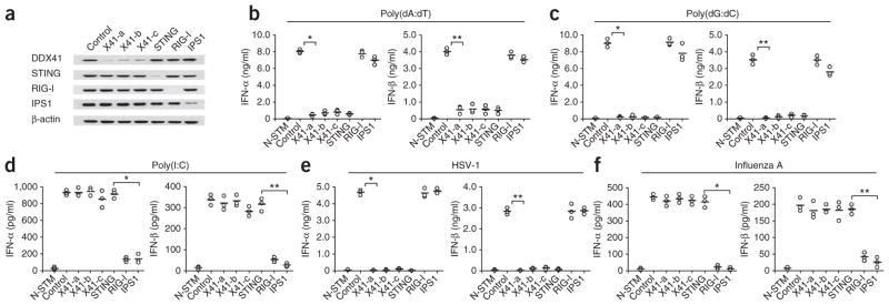
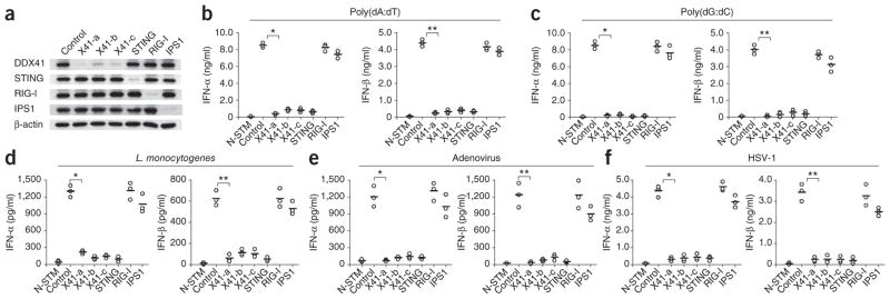
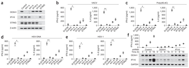
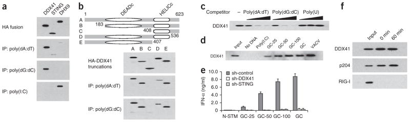
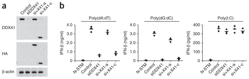
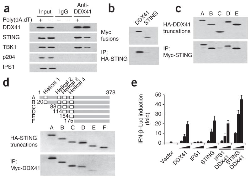
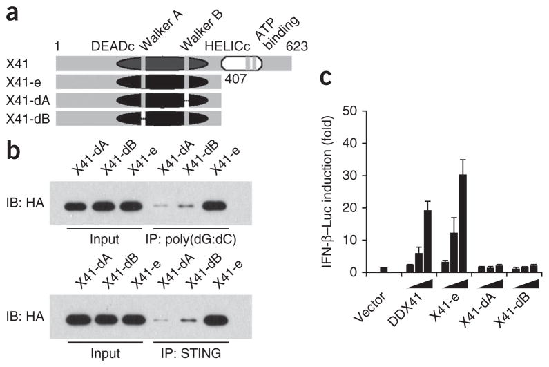
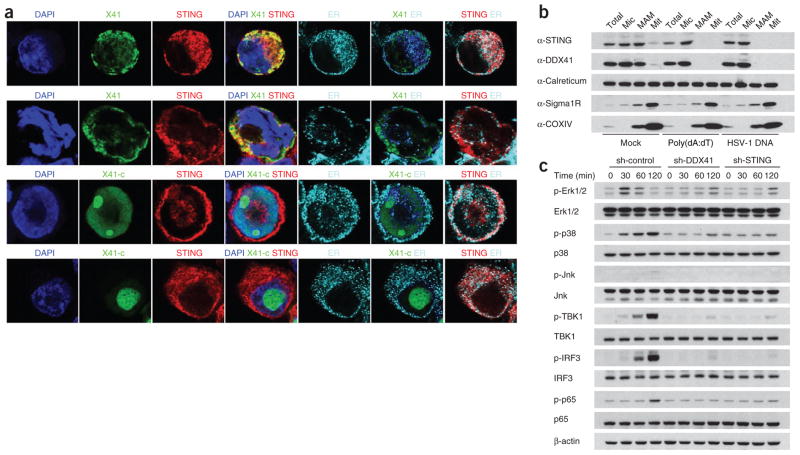
The recognition of pathogenic DNA is important to the initiation of antiviral responses. Here we report the identification of DDX41, a member of the DEXDc family of helicases, as an intracellular DNA sensor in myeloid dendritic cells (mDCs). Knockdown of DDX41 expression by short hairpin RNA blocked the ability of mDCs to mount type I interferon and cytokine responses to DNA and DNA viruses. Overexpression of both DDX41 and the membrane-associated adaptor STING together had a synergistic effect in promoting Ifnb promoter activity. DDX41 bound both DNA and STING and localized together with STING in the cytosol. Knockdown of DDX41 expression blocked activation of the mitogen-activated protein kinase TBK1 and the transcription factors NF-κB and IRF3 by B-form DNA. Our results suggest that DDX41 is an additional DNA sensor that depends on STING to sense pathogenic DNA.


Conflict of interest statement

COMPETING FINANCIAL INTERESTS

The authors declare no competing financial interests.

Figures

Figure 1
Figure 1
DDX41 senses cytosolic DNA in mDCs. (a) Immunoblot analysis of the knockdown efficiency of nontargeting scrambled shRNA (Control) or shRNA targeting mRNA encoding DDX41 (three shRNAs: X41-a, X41-b and X41-c), STING, RIG-I or IPS1 (above lanes) in D2SC mDCs; β-actin (bottom) serves as a loading control throughout. (bf) ELISA of IFN-α and IFN-β in D2SC cells either treated with scrambled shRNA and left unstimulated (N-STM) or treated with scrambled shRNA (Control) or shRNA targeting mRNA encoding DDX41, STING, RIG-I or IPS1 (as in a; horizontal axis), then stimulated for 16 h with poly(dA:dT) (1 μg/ml; b), poly(dG:dC) (1 μg/ml; c), poly(I:C) (2.5 μg/ml; d), HSV-1 (multiplicity of infection (MOI), 10; e) or influenza A virus (MOI, 10; f). Each symbol represents the result of one experiment; small horizontal lines indicate the average. (b) *P < 1.5 × 10⁻⁶ and **P < 3.5 × 10⁻⁵; (c) *P < 8.3 × 10⁻⁶ and **P < 2.8 × 10⁻⁵; (d) *P < 5.2 × 10⁻⁵ and **P < 9.5 × 10⁻⁵; (e) *P < 8.10 × 10⁻⁷ and **P < 6.0 × 10⁻⁶; and (f) *P < 3.2 × 10⁻⁵ and **P < 2.0 × 10⁻⁴ (Student’s t-test). Data are from at least three independent experiments.
Figure 2
Figure 2
Knockdown of DDX41 or STING in BMDCs abolishes their cytokine responses to DNA alone and DNA viruses. (a) Immunoblot analysis of the knockdown efficiency of shRNA (as in Fig. 1a) in BMDCs. (bf) ELISA (as in Fig. 1a) of IFN-α and IFN-β in DCs either treated with scrambled shRNA and left unstimulated or treated with scrambled shRNA or shRNA (as in a), then stimulated for 16 h with poly(dA:dT) (1 μg/ml; b), poly(dG:dC) (1 μg/ml; c), L. monocytogenes (20 colony-forming units; d), adenovirus (MOI, 10; e) or HSV-1 (MOI, 10; f). (b) *P < 8.3 × 10⁻⁷ and **P < 4.0 × 10⁻⁶; (c) *P < 6.0 × 10⁻⁶ and **P < 1.8 × 10⁻⁵; (d) *P < 5.7 × 10⁻⁵ and **P < 2.5 × 10⁻⁴; (e) *P < 4.4 × 10⁻⁴ and **P < 6.4 × 10⁻⁴; and (f) *P < 3.4 × 10⁻⁵ and **P < 1.9 × 10⁻⁴ (Student’s t-test). Data are from at least three independent experiments.
Figure 3
Figure 3
Knockdown of DDX41 or STING in THP-1 cells abolishes their cytokine responses to DNA or HSV-1. (a) Immunoblot analysis of the knockdown efficiency of scrambled shRNA or shRNA targeting mRNA encoding DDX41 (two shRNAs: X41-a and X41-b), IFI16 (two shRNAs: IFI16-a and IFI16-b) or STING (above lanes ) in THP-1 cells. (be) ELISA (as in Fig. 1a) of IFN-β and IL-6 in THP-1 cells either treated with nontargeting scrambled shRNA and left unstimulated or treated with shRNA (as in a), then stimulated for 16 h with vaccinia virus (VACV) DNA (1 μg/ml; b), poly(dG:dC) (1 μg/ml; c), HSV DNA (1 μg/ml; d) or HSV-1 (MOI, 10; e). (b) *P < 4.0 × 10⁻⁶ and **P < 2.0 × 10⁻⁵; (c) *P < 3.6 × 10⁻⁵ and **P < 2.2 × 10⁻⁵; (d) *P < 9.3 × 10⁻⁵ and **P < 5.10 × 10⁻⁵; (e) *P < 7.0 × 10⁻⁵ and **P < 7.8 × 10⁻⁵ (Student’s t-test). (f) ELISA of IFN-β (as in Fig. 1a; top) and immunoblot analysis of the expression of DDX41 and IFI16 (below) in THP-1 cells treated with nontargeting shRNA or shRNA targeting DDX41 or IFI16 and stimulated for 0–16 h with poly(dA:dT) (1 μg/ml). GAPDH (glyceraldehyde phosphate dehydrogenase) serves as a loading control (bottom). Data are from at least three independent experiments.
Figure 4
Figure 4
DDX41 interacts with DNA but not with RNA. (a) Immunoblot analysis of immunoprecipitation (IP) assays of purified HA-tagged DDX41, STING or DHX9 incubated with biotinylated poly(dA:dT), poly(dG:dC) or poly(I:C), probed with anti-HA. (b) Immunoblot analysis of immunoprecipitation assays of purified HA-tagged full-length DDX41 (A) and serial truncations of DDX41 (B–E) incubated individually with biotinylated DNA, probed with anti-HA. Top, full-length and serial truncations of DDX41. DEADc, Asp-Glu-Ala-Asp motif; HELICc, helicase C-terminal domain; numbers indicate positions of amino acids. (c) Immunoblot analysis of nucleic acid–immunoprecipitation competition assays of increasing concentrations of DNA or poly(U) (0.5, 5 or 50 μg/ml; wedges) or no nucleic acid (−) added to a mixture of HA-tagged DDX41 plus biotinylated poly(dA:dT), probed with anti-HA. (d) Immunoblot analysis of immunoprecipitation assays of purified HA-tagged DDX41 incubated with avidin beads alone (No DNA) or with various biotinylated RNA or DNA substrates plus avidin beads, probed with anti-HA. Input, 10% of the purified HA-tagged DDX41; GC-25, GC-50 or GC-100, poly(dG:dC) 25, 50 or 100 nucleotides in length; GC, full-length poly(dG:dC); VACV, vaccinia virus. (e) ELISA of IFN-α in D2SC cells treated with control shRNA (sh-control) or shRNA targeting DDX41 (sh-DDX41) or STING (sh-STING) and left unstimulated (N-STM) or stimulated with poly(dG:dC) of various lengths (as in d). (f) Immunoblot analysis of endogenous DDX41, p204 and RIG-I in D2SC cells incubated for 0 or 60 min with biotinylated poly(dA:dT). Input, 10% of the D2SC lysate. Data are representative of three independent experiments (mean and s.d. in e).
Figure 5
Figure 5
Recombinant DDX41 can ‘rescue’ the defect in DNA-activated production of interferon caused by knockdown of DDX41 via siRNA. (a) Immunoblot analysis of endogenous DDX41 in D2SC mDCs (Control) or of recombinant HA-tagged full-length or truncated DDX41 in D2SC mDCs with selective knockdown of endogenous DDX41 alone (siDDX41) or along with expression of full-length DDX41 (si+X41-a) or DDX41 with deletion of the DNA-binding domain (si+X41-c). (b) ELISA (as in Fig. 1a) of IFN-β in D2SC mDCs either left untreated and unstimulated (N-STM) or treated as in a and stimulated for 16 h with poly(dA:dT) (1 μg/ml), poly(dG:dC) (1 μg/ml) or poly(I:C) (2.5 μg/ml). Data are from at least three independent experiments.
Figure 6
Figure 6
Interaction of DDX41 with STING. (a) Immunoblot analysis of proteins (left margin) precipitated with anti-DDX41 or immunoglobulin G (IgG; control) from whole-cell lysates of D2SC cells left unstimulated (−) or stimulated with poly(dA:dT) (+). Input, 10% of the D2SC cells lysate. (b) Immunoblot analysis of immunoprecipitation assays of purified HA-tagged STING incubated with Myc-tagged DDX41 or STING, probed with anti-Myc. (c) Immunoblot analysis of immunoprecipitation assays of purified HA-tagged full-length or truncated DDX41 (as in Fig. 4b) incubated with Myc-tagged STING, probed with anti-HA. (d) Immunoblot analysis of purified HA-tagged full-length STING (A) or truncated STING (B–F), probed with anti-HA (middle), and immunoblot analysis of immunoprecipitation assays of HA-tagged STING (as above) incubated with Myc-tagged DDX41, probed with anti-HA (bottom). Top, full-length STING (A) and serial truncations of STING (B–F); numbers indicate positions of amino acids. (e) Activation of the Ifnb promoter in mouse L929 cells transfected with an IFN-β luciferase reporter (IFN-β–Luc; 100 ng) plus increasing concentrations (20, 100 or 200 ng; wedges) of expression vectors for DDX41, IPS1 or STING individually; or expression vector for DDX41 (20 ng; solid bar) together with increasing concentrations (20, 100 or 200 ng; wedges) of expression vectors for IPS1 or STING. Results are presented relative to those of cells transfected with empty vector alone (Vector). Data are representative of three independent experiments (mean and s.d. in e).
Figure 7
Figure 7
The Walker motifs in DDX41 are essential for sensing DNA. (a) Full-length DDX41 (X41) and serial deletion mutants of DDX41 lacking the HELICc domain (X41-e), both the Walker A motif and the HELICc domain (X41-dA) or both the Walker B motif and the HELICc domain (X41-dM). (b) Immunoblot analysis of immunoprecipitation assays of purified HA-tagged DDX41 (as in a) incubated with biotinylated poly(dG:dC) followed by addition of avidin beads (top), or with Myc-tagged STING, followed by the addition of anti-Myc beads (bottom), probed with anti-HA. Input, 10% of the purified HA-tagged DDX41. (c) Activation of the Ifnb promoter in L929 cells transfected with the IFN-β luciferase reporter (100 ng) plus increasing concentrations (20, 100 or 200 ng; wedges) of expression vectors for DDX41 (as in a); a renilla luciferase reporter (2 ng) was transfected simultaneously as an internal control. Results are presented relative to those of cells transfected with empty vector alone (Vector). Data are representative of three independent experiments (mean and s.d. in c).
Figure 8
Figure 8
STING is the key adaptor for DDX41 signaling. (a) Confocal microscopy of HEK293T cells transfected with expression plasmid for HA-tagged STING, Myc-tagged DDX41 (X41) and/or Myc-tagged DDX41 with truncation of the C terminus (X41-c), then left unstimulated (top and third rows) or stimulated for 4 h with poly(dA:dT) (second and bottom rows). Nuclei are stained with the DNA-intercalating dye DAPI; staining of calreticulin serves as a marker of the endoplasmic reticulum (ER). Original magnification, ×100. Data are representative of three independent experiments. (b) Immunoblot analysis of the fractionation of unstimulated D2SC cells (Mock) or D2SC cells stimulated with poly(dA:dT) (1 μg/ml) or HSV-1 DNA (1 μg/ml), probed with anti- STING, anti-DDX41, anti-calreticulin (to detect the endoplasmic reticulum), anti-Sigma1R (to detect the mitochondria-associated ER membrane (MAM)) or anti-COXIV (to detect mitochondria (Mit)). Total, 15% of the D2SC lysate; Mic, microsome. Data are representative of three experiments. (c) Immunoblot analysis of phosphorylated (p-) and total Erk1/2, p38, Jnk, TBK1, IRF3 and p65 in lysates of BMDCs treated with shRNA (as in Fig. 4e) and stimulated for 0–120 min with poly(dA:dT) (1 μg/ml). Data are representative of three experiments.

Comment in

  • STING-dependent signaling.
    Barber GN. Barber GN. Nat Immunol. 2011 Sep 20;12(10):929-30. doi: 10.1038/ni.2118. Nat Immunol. 2011. PMID: 21934672

Similar articles

Cited by

References

    1. Iwasaki A, Medzhitov R. Toll-like receptor control of the adaptive immune responses. Nat Immunol. 2004;5:987–995. - PubMed
    1. Takeuchi O, Akira S. Recognition of viruses by innate immunity. Immunol Rev. 2007;220:214–224. - PubMed
    1. Blasius AL, Beutler B. Intracellular toll-like receptors. Immunity. 2010;32:305–315. - PubMed
    1. Kato H, et al. Differential roles of MDA5 and RIG-I helicases in the recognition of RNA viruses. Nature. 2006;441:101–105. - PubMed
    1. Myong S, et al. Cytosolic viral sensor RIG-I is a 5′-triphosphate-dependent translocase on double-stranded RNA. Science. 2009;323:1070–1074. - PMC - PubMed