. 2008:2:275-87.
doi: 10.4137/cmo.s615. Epub 2008 Mar 27.

Multiple Myeloma Includes Phenotypically Defined Subsets of Clonotypic CD20+ B Cells that Persist During Treatment with Rituximab

Affiliations

Multiple Myeloma Includes Phenotypically Defined Subsets of Clonotypic CD20+ B Cells that Persist During Treatment with Rituximab

Linda M Pilarski et al. Clin Med Oncol. 2008.

Abstract

Potential progenitor B cell compartments in multiple myeloma (MM) are clinically important. MM B cells and some circulating MM plasma cells express CD20, predicting their clearance by treatment with anti-CD20. Here we describe two types of clonotypic CD20+ B cell in peripheral blood of myeloma patients, identified by their expression of CD19 and CD20 epitopes, their expression of CD45RA and their light scatter properties. Thus, the circulating component of the MM clone includes at least two distinct CD19+ CD20+ B cell compartments, as well as CD138+ CD20+ plasma cells. To determine whether either or both B cell subsets and the CD20+ plasma cell subset were depleted by anti-CD20 therapy, they were evaluated before, during and after treatment of patients with rituximab (anti-CD20), followed by quantifying B cell subsets over a 5 month period during and after treatment. Overall, all three types of circulating B lineage cells persist despite treatment with rituximab. The inability of rituximab to prolong survival in MM may result from this failure to deplete CD20+ B and plasma cells in MM.

Keywords: B cell depletion by rituximab; B lineage cells; CD19 epitopes; CD20 epitopes; multiple myeloma.


Figures

Figure 1
Figure 1. Expression of CD19 Epitopes on B cell populations in multiple myeloma and in healthy donors: Absence of the Leu12 epitope on most MM B cells
PBMC from a representative healthy donor (top row of panels) and a representative MM patient (bottom row) were stained in two color immunofluoresence with CD3-PE and three types of CD19-FITC, B4, FMC63 and Leu12 as indicated in methods. The mean values for each antibody are given in Table 1. Files were ungated. Sorted B cells stained as shown here, and sorted within the analysis gates shown in this figure have been shown to express intracellular immunoglobulin, CD19 transcripts and IgH transcripts (Pilarski, Giannakopoulos, Szczepek, Masellis, Mant, and Belch, 2000a; Szczepek, Bergsagel, Axelsson, Brown, Belch, and Pilarski, 1997) as well as clonotypic IgH VDJ (Pilarski, Giannakopoulos, Szczepek, Masellis, Mant, and Belch, 2000a; Szczepek, Bergsagel, Axelsson, Brown, Belch, and Pilarski, 1997; Szczepek, Seeberger, Wizniak, Mant, Belch, and Pilarski, 1998).
Figure 2
Figure 2. Two distinct populations of B cells in MM patients
Respresentative staining of PBMC from a healthy donor and an MM patient in two color immunofluorescence with Leu12-PE and FMC63-FITC, showing two distinct populations of B cells in MM patients (CD19+ Leu12− and CD19+ Leu12+) but only one in healthy donors (CD19+Leu12+). Previous work has shown that the Leu12− populations detected by B4 and FMC63 are B cells and express clonotypic transcripts (Pilarski, Giannakopoulos, Szczepek, Masellis, Mant, and Belch 2000a; Pilarski, Szczepek, and Belch 1997; Szczepek, Seeberger, Wizniak, Mant, Belch, and Pilarski 1998; Szczepek, Bergsagel, Axelsson, Brown, Belch, and Pilarski 1997).
Figure 3
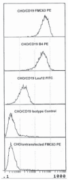
Figure 3. Expression of CD19 epitopes on CD19 transfectants
To confirm that all three antibodies detected CD19 epitopes, FMC63, B4 and Leu12 antibodies were used to stain CD19 transfectants. The transfectants were shown to express the CD19 epitope as detected by binding of each antibody. Appropriate isotype controls were performed for each anti-CD19 antibody. The observed staining with Leu12 indicates that in transfectants, unlike MM cells but similar to B cells from healthy donors, the Leu12 epitope is exposed.
Figure 4
Figure 4. Neuraminidase treatment reveals Leu12 epitopes on MM B cells
MM PBMC were treated with neuraminidase (solid line) or left untreated (dashed line) as described in methods, followed by staining with Leu12-PE. The marker bar indicates staining above negative control values. The numerical value above the marker bar reports the number of Leu12+ cells after neuraminidase treatment. The Leu12bright peak between 102 and 103 represents expression that is independent of neuraminidase treatment (dotted line), while the lower intensity peak represents Leu12 epitopes revealed by neuraminidase (solid line). PBMC were also stained before and after treatment with fluorescent conjugates of FMC63, B4, CD20 mAbs B1 and rituximab; the staining profiles were comparable before and after treatment for all antibodies except Leu12.
Figure 5
Figure 5. Leu12+ B cells have low forward and side scatter, in contrast to FMC63+ B cells which include a subpopulation with high forward and side scatter
MM PBMC were stained with Leu12 or FMC63. Files were gated on the CD19+ population for each antibody: Leu12+ = 3.3% of PBMC and FMC63+ = 29% OF PBMC. B4+ B cells had the same scatter profile as did FMC63+ B cells. B1+ or rituximab+ B cells also have a scatter profile comparable to that of the FMC63+ B cells. The vertical axis represents side scatter (SSC) and the horizontal axis represents forward scatter (FSC). The horizontal axes from left to right represent increasing extent of forward scatter. The left panel shows a single population of Leu12+ B cells having low side and forward scatter. The right panel shows the two subpopulations distinguished by FMC63 (FMC63+ high side or forward scatter, and FMC63+ low side or forward scatter).
Figure 6
Figure 6. Phenotypic analysis of CD19, CD20 and CD45RA identify two distinct populations of CD19+20+ B cells
Representative MM PBMC were stained with anti-CD19-FITC (horizontal axis) and either CD20-PE vertical axis, top row) or CD45RA-PE (vertical axis, bottom row). The boxes indicate subopulations defined by this analysis. The left top panel shows FMC63+ CD20Moderate, and FMC63+CD20bright subpopulations, and the left bottom panel shows FMC63+CD45RAbright and FMC63+CD45RAdim populations. The right panels show a single population identified by Leu12.
Figure 7
Figure 7. A proportion of Leu12+ MM B cells are clonotypic
FMC63+Leu12− (monocytoid gates) and FMC63+Leu12+ (lymphocyte gates) B cells were sorted and analyzed for clonotypic IgH VDJ transcripts using in situ RT-PCR. Leu12− monocytoid B cells were sorted from PBMC of 12 MM patients. Leu12+ lymphocyic B cells were sorted from PBMC of 15 MM patients. Leu12 negative B cells included significantly more clonotypic B cells than did Leu12+ B cells (p = .003). One of the patients analyzed for clonotypic Leu12+ B cells was in a terminal stage of disease.

Similar articles

Cited by

References

    1. Bakkus MH, Van Riet C, Van Camp I, Thielemann B. Evidence that the clonogenic cell in multiple myeloma originates from a pre-switched but somatically mutated B. cell. Brit J Hematol. 87:68–74. - PubMed
    1. Bergsagel PL, Masellis S, Szczepek A, Mant MJ, Belch AR, Pilarski LM. In multiple myeloma, clonotypic B. lymphocytes are detectable among CD19+ peripheral blood cells expressing CD38, CD56 and monotypic immunoglobulin light chain. Blood. 1995;85:436–47. - PubMed
    1. Bonavida B. Rituximab-induced inhibition of antiapoptotic cell survival pathways: implications in chemo/immunoresistance, rituximab unresponsiveness, prognostic and novel therapeutic interventions. Oncogene. 2007;26(25):3629–36. - PubMed
    1. Byrd JC, Kitada S, Flinn IW, Aron JL, Pearson M, Lucas D, Reed JC. The mechanism of tumor cell clearance by rituximab in vivo in patients with B.-cell chronic lymphocytic leukemia: evidence of caspase activation and apoptosis induction. Blood. 2002;99(3):1038–43. - PubMed
    1. Cone L, Uhr JW. Immunological deficiency disorders associated with chronic lymphocytic leukemia and multiple myeloma. J Clin Invest. 1964;43:2241–8. - PMC - PubMed

LinkOut - more resources