. 2012 Mar;11(1):300-10.
doi: 10.1007/s12311-011-0311-0.

An fMRI investigation of cerebellar function during verbal working memory in methadone maintenance patients

Affiliations

An fMRI investigation of cerebellar function during verbal working memory in methadone maintenance patients

Cherie L Marvel et al. Cerebellum. 2012 Mar.

Abstract

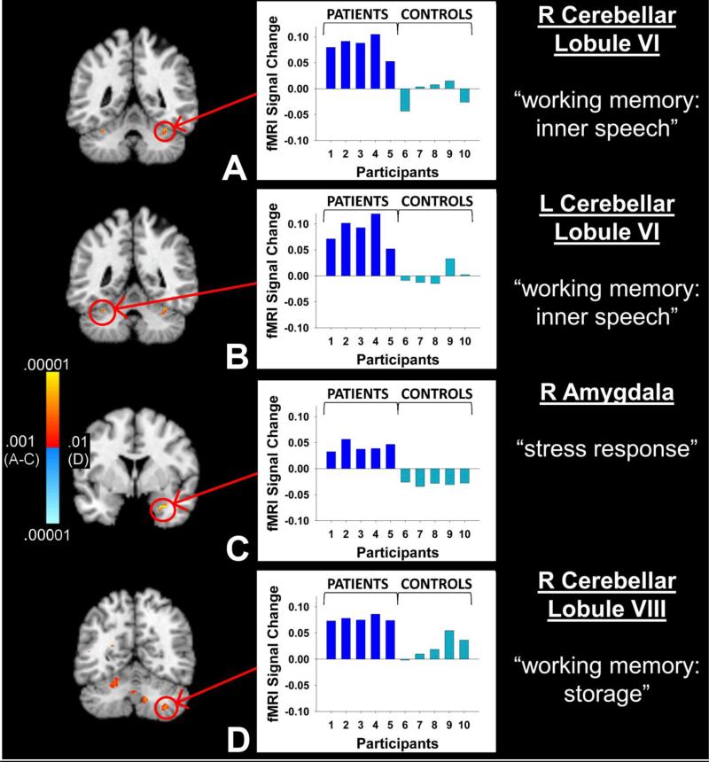
Working memory is impaired in opioid-dependent individuals, yet the neural underpinnings of working memory in this population are largely unknown. Previous studies in healthy adults have demonstrated that working memory is supported by a network of brain regions that includes a cerebro-cerebellar circuit. The cerebellum, in particular, may be important for inner speech mechanisms that assist verbal working memory. This study used functional magnetic resonance imaging to examine brain activity associated with working memory in five opioid-dependent, methadone-maintained patients and five matched, healthy controls. An item recognition task was administered in two conditions: (1) a low working memory load "match" condition in which participants determined whether target letters presented at the beginning of the trial matched a probe item, and (2) a high working memory load "manipulation" condition in which participants counted two alphabetical letters forward of each of the targets and determined whether either of these new items matched a probe item. Response times and accuracy scores were not significantly different between the groups. FMRI analyses indicated that, in association with higher working memory load ("manipulation" condition), the patient group exhibited hyperactivity in the superior and inferior cerebellum and amygdala relative to that of controls. At a more liberal statistical threshold, patients exhibited hypoactivity in the left prefrontal and medial frontal/pre-SMA regions. These results indicate that verbal working memory in opioid-dependent individuals involves a disrupted cerebro-cerebellar circuit and shed light on the neuroanatomical basis of working memory impairments in this population.


Figures

Figure 1
Figure 1
A verbal working memory task was given under two conditions. a) In the match condition, participants encoded 1 or 2 targets (encoding phase). Note that in the example, 2 targets are shown, but on half the trials, only 1 target was shown. Then, participants silently rehearsed these targets across a delay (delay phase). Finally, at the presentation of a letter probe, subjects indicated whether the probe matched either of the targets (retrieval phase). b) In the manipulation condition, the encoding phase was identical to that in the match condition. However, in the delay phase, instead of simply rehearsing the targets, subjects counted two alphabetical letters forward of each. Then they rehearsed these newly identified targets. In the retrieval phase, subjects indicated whether the probe matched either of the newly identified targets (rather than the original targets). In both figures, the box wave indicates when stimuli were visible on the screen.
Figure 2
Figure 2
Behavioral results. a) Mean accuracy for all trials as a function of condition (match vs. manipulation), target load (1 vs. 2 targets) and group (controls vs. patients). Significant accuracy effects were found for condition and target load, as well as for an interaction of condition × target load (i.e., low accuracy for the 2-item target load in the manipulation condition). b) Mean response times for correct responses as a function of condition, target load, and group. Significant response time effects were found for condition (slower on the manipulation condition) and target load (slower on 2-item trials). Error bars of 1 standard error are plotted for each bar in the graphs. Manip = Manipulation condition.
Figure 3
Figure 3
Group differences in fMRI signal contrast for the manipulation minus match conditions. Left: color coded group activation differences have been overlaid onto a standard MNI anatomical image, using neurological orientation. Positive differences (patients > controls) are shown in yellow/red and negative differences (controls > patients) are shown in blue; p < .001 - .00001 for slices A-C and p < .01 - .00001 for slice D to improve visualization of the sub-threshold cluster size. [Note that decreasing the threshold in D revealed additional activations which were not discussed here.] The primary activation of interest within each slice is circled in red. Middle: individual fMRI signal contrast values for patients and controls are graphed separately for each activation of interest. Right: the names of the brain regions corresponding to the activations of interest are listed along with the proposed functional contributions of each region to the task. L = left, R = right.
Figure 4
Figure 4
Consistency of group activation differences across studies of addiction and working memory. a) Location of increased cerebellar activations associated with high verbal working memory load as reported in four separate neuroimaging studies: 1) orange = opioid-dependent patients in the current study, 2) red = individuals with alcohol dependence [19], 3) green = individuals with cocaine dependence [20], and 4) yellow = healthy, young adults performing the same task that was administered in the current study (Marvel & Desmond, submitted). Peak activations in the comparative studies were obtained directly from the publications and converted to MNI coordinates using local software [27, 51]. Foci were plotted manually by CLM using Paint.NET software on coronal section y = -58, which was the mean y-axis location across the four studies, and overlaid onto the atlas section developed by Schmahmann et al. [28], shown in neurological orientation. Superior cerebellar activations were consistent across the studies. In addition, increased right inferior cerebellar activity was observed in the patients of the current study (orange) and in healthy, young adults (yellow). b) Location of group activation differences in the left prefrontal lobe (BA 9) reported in the comparative studies listed in part (a), plus a fifth study, shown in blue, that represents a study of opioid-dependence [21]. Activation locations were converted from Talairach to MNI coordinates as described in 4a. Foci were plotted using Tor Wager's Meta Analysis Toolbox http://www.columbia.edu/cu/psychology/tor/meta-analysis.html. c) Location of group activation differences in the medial frontal gyrus (BA 6/pre-SMA), shown in the right hemisphere, with axes as in 4b. The orange markers in 4b and 4c represent hypoactivations by the patients in the current study, at the sub-threshold level of p < .05.

References

    1. Baddeley A. Working memory. Science. 1992;255(5044):556–9. - PubMed
    1. Ackermann H, Mathiak K, Ivry RB. Temporal organization of “internal speech” as a basis for cerebellar modulation of cognitive functions. Behav Cogn Neurosci Rev. 2004;3(1):14–22. - PubMed
    1. Levy BA. Role of Articulation in Auditory and Visual Short-Term Memory. Journal of Verbal Learning and Verbal Behavior. 1971:10123–132.
    1. Murray DJ. The effect of white noise upon the recall of vocalized lists. Can J Psychol. 1965;19(4):333–45. - PubMed
    1. Marvel CL, Desmond JE. Functional topography of the cerebellum in verbal working memory. Neuropsychol Rev. 2010;20(3):271–9. - PMC - PubMed

Publication types

MeSH terms