

Case Reports
doi: 10.1007/s12105-011-0294-7.
Epub 2011 Sep 3.
Clinical pathologic conference case 6: infantile myofibroma
Affiliations
- PMID: 21892764
- PMCID: PMC3173535
- DOI: 10.1007/s12105-011-0294-7
Item in Clipboard
Case Reports
Clinical pathologic conference case 6: infantile myofibroma
Head Neck Pathol.
2011 Sep.
No abstract available
Figures
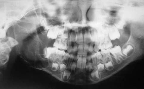
Panoramic radiography of a 5-year-old girl, showing a large well-delimited multilocular radiolucent lesion involving the right angle and ascending ramus of the mandible
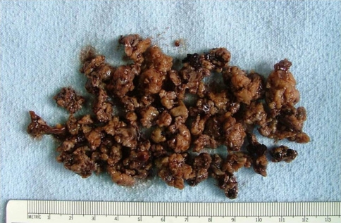
Macroscopic aspects of the biopsy formed of multiple brownish elastic fragments
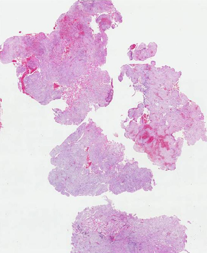
General microscopic view at low magnification, showing multiple fragments formed by fibrous tissue with areas of hemorrhage (H&E, 40×)
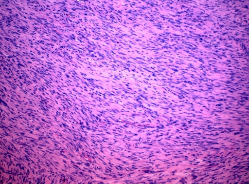
Fascicles of spindle cells, hyperchromatic nuclei, and indistinct cytoplasm, permeated by some hypocellular areas (H&E, 100×)
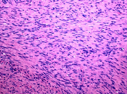
Higher magnification of variable densities of spindle cells in longitudinal and transversal sections, exhibiting myofibroblastic morphology and eosinophilic background (H&E, 200×)
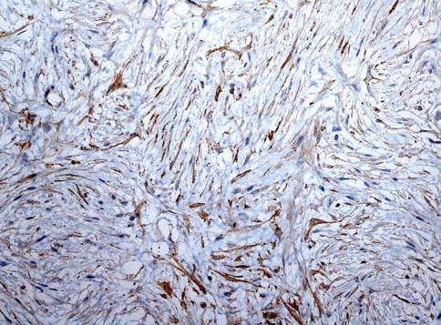
Tumor cells were strongly positive for smooth muscle actin (IHC, 200×)
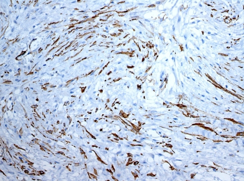
Most of the tumor cells were positive for muscle-specific actin (HHF-35; IHC, 200×)
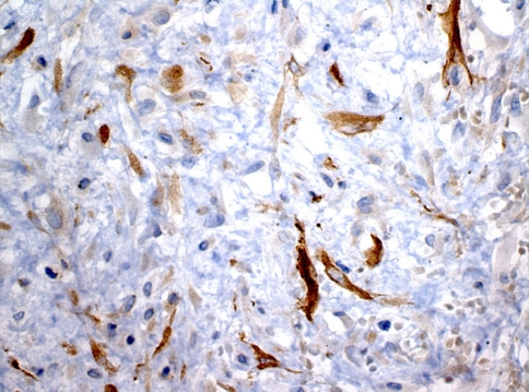
Many tumor cells were also strongly positive for calponin (IHC, 400×)
References
Publication types
MeSH terms
LinkOut - more resources
Full Text Sources
