. 2011 Sep 5:5:433.
doi: 10.1186/1752-1947-5-433.

Cardiac tamponade and paroxysmal third-degree atrioventricular block revealing a primary cardiac non-Hodgkin large B-cell lymphoma of the right ventricle: a case report

Affiliations

Cardiac tamponade and paroxysmal third-degree atrioventricular block revealing a primary cardiac non-Hodgkin large B-cell lymphoma of the right ventricle: a case report

Zied Frikha et al. J Med Case Rep. .

Abstract

Introduction: Primary cardiac lymphoma is rare.

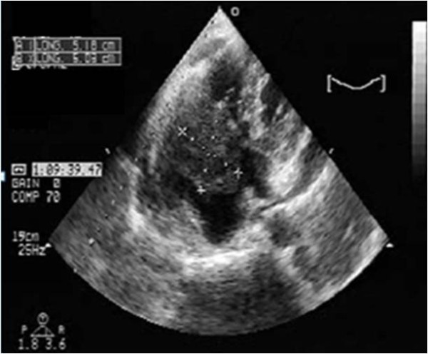
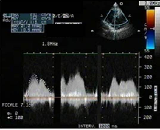
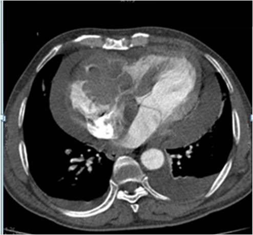
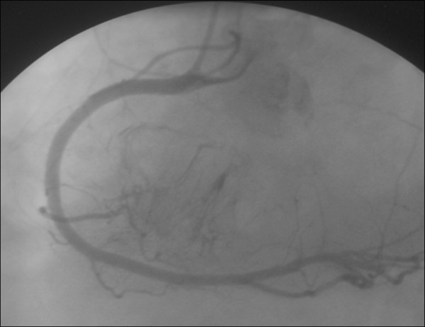
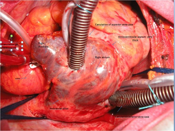
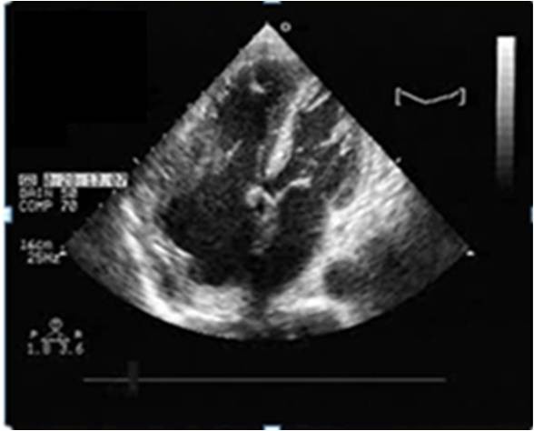
Case presentation: We report the case of a 64-year-old non-immunodeficient Caucasian man, with cardiac tamponade and paroxysmal third-degree atrioventricular block. Echocardiography revealed the presence of a large pericardial effusion with signs of tamponade and a right ventricular mass was suspected. Scanner investigations clarified the sites, extension and anatomic details of myocardial and pericardial infiltration. Surgical resection was performed due to the rapid impairment of his cardiac function. Analysis of the pericardial fluid and histology confirmed the diagnosis of non-Hodgkin large B-cell lymphoma. He was treated with chemotherapy.

Conclusion: The prognosis remains poor for this type of tumor due to delays in diagnosis and the importance of the site of disease.


Figures

Figure 1
Figure 1
Transthoracic echocardiography four chamber view showing a pericardial effusion and a large mass. The mass measured 5.5 cm× 5 cm in the right ventricle and was attached to the tricuspid valve creating a tricuspid stenosis. The tumor has spread over the right atrium.
Figure 2
Figure 2
Continuous wave Doppler. The tumor created a hemodynamic tricuspid stenosis which is a sign of high right ventricle inflow velocities.
Figure 3
Figure 3
Coronary angiography showing an accentuation of myocardial blush.
Figure 4
Figure 4
Computed tomography scan showing the presence of a right heart tumor developing on both sides of the tricuspid valve.
Figure 5
Figure 5
Macroscopic aspect of the tumor. The tumor is infiltrating the right atrium, the atrioventricular septum and the proximal side of the right ventricle.
Figure 6
Figure 6
Macroscopic aspect of the tumor which is infiltrating the right atrium.
Figure 7
Figure 7
A transthoracic echocardiography four chamber view showing a reduction of the size of the mass.

Similar articles

Cited by

References

    1. Mikdame M, Ennebi K, Bahrouch L, Benyass A, Dreyfus F, Touloune F. Localisations cardiaques du lymphome non hodgkinien: à propos de quatre cas. Rev Med Interne. 2003;24:459–463. doi: 10.1016/S0248-8663(03)00144-9. - DOI - PubMed
    1. Fuzellier JF, Saade YA, Torossian PF, Baehrel B. Primary cardiac lymphoma: Diagnosis and treatment. Arch Mal Coeur Vaiss. 2005;98:875–880. - PubMed
    1. Sutcliffe SB, Gospodarowicz MK. In: The Lymphomas. Canellos GP, Lister TA, Sklar JL, editor. Philadelphia: WB Saunders Company; 1998. Primary extranodal lymphomas; pp. 449–479.
    1. Quigley MM, Schwartzman E, Boswell PD, Christensen RL, Gleason LA, Sharpe RW, D'Amato TA. A unique atrial primary cardiac lymphoma mimicking myxoma presenting with embolic stroke: A case report. Blood. 2003;101:4708–4710. doi: 10.1182/blood-2002-08-2550. - DOI - PubMed
    1. Ceresoli GL, Ferreri AJM, Bucci E, Ripa C, Ponzoni M, Villa E. Primary cardiac lymphoma in immunocompetent patients - diagnostic and therapeutic management. Cancer. 1997;80:1497–1506. doi: 10.1002/(SICI)1097-0142(19971015)80:8<1497::AID-CNCR18>3.0.CO;2-0. - DOI - PubMed

LinkOut - more resources