Review
. 2011 Nov;63(14-15):1300-31.
doi: 10.1016/j.addr.2011.08.002. Epub 2011 Aug 26.

Magnetically enhanced nucleic acid delivery. Ten years of magnetofection-progress and prospects

Affiliations
Review

Magnetically enhanced nucleic acid delivery. Ten years of magnetofection-progress and prospects

Christian Plank et al. Adv Drug Deliv Rev. 2011 Nov.

Abstract

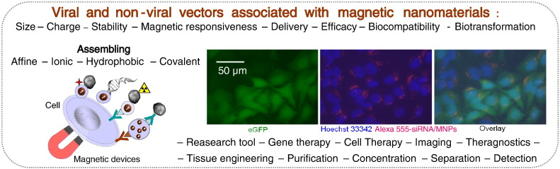
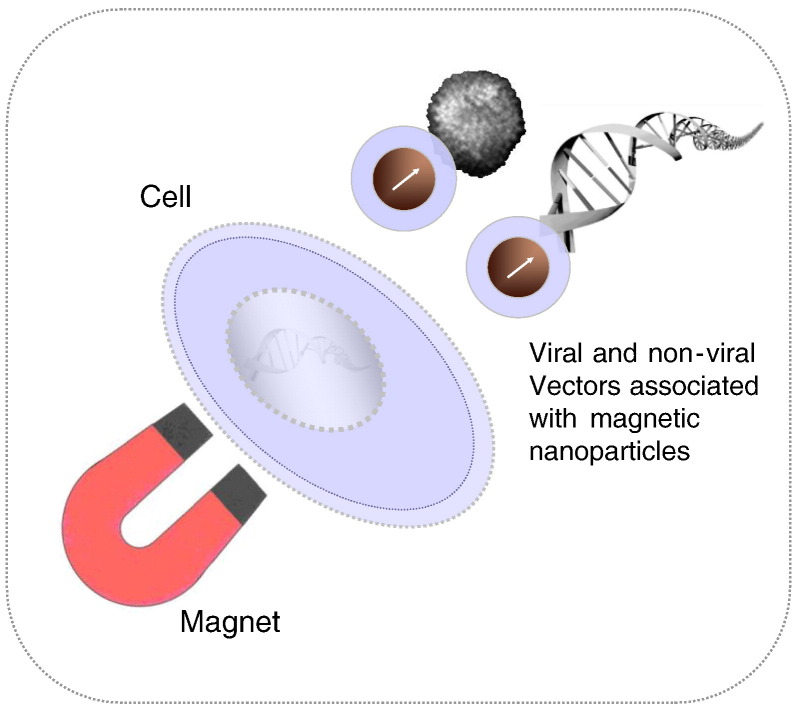
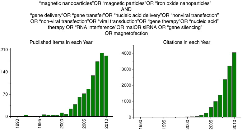
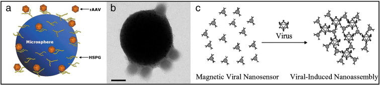
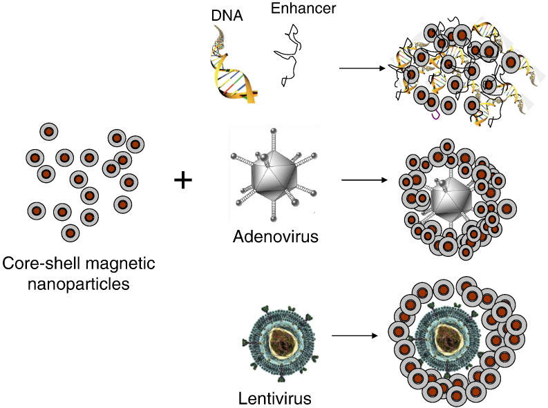
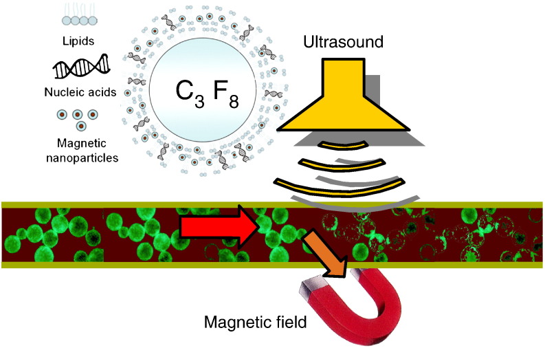
Nucleic acids carry the building plans of living systems. As such, they can be exploited to make cells produce a desired protein, or to shut down the expression of endogenous genes or even to repair defective genes. Hence, nucleic acids are unique substances for research and therapy. To exploit their potential, they need to be delivered into cells which can be a challenging task in many respects. During the last decade, nanomagnetic methods for delivering and targeting nucleic acids have been developed, methods which are often referred to as magnetofection. In this review we summarize the progress and achievements in this field of research. We discuss magnetic formulations of vectors for nucleic acid delivery and their characterization, mechanisms of magnetofection, and the application of magnetofection in viral and nonviral nucleic acid delivery in cell culture and in animal models. We summarize results that have been obtained with using magnetofection in basic research and in preclinical animal models. Finally, we describe some of our recent work and end with some conclusions and perspectives.


Figures

None
Graphical abstract
Fig. 1
Fig. 1
Principle of magnetofection: viral or non-viral gene delivery vectors are associated with magnetic nanoparticles. Magnetic force directs vectors towards target cells resulting in rapid and highly efficient nucleic acid delivery.
Fig. 2
Fig. 2
Results of the search in the ISI Web of Science web site as defined in the figure.
Fig. 3
Fig. 3
Assembling of the virus and magnetic particles due to specific ligand–ligand interactions. (a) Schematic model of microsphere-conjugated rAAV. (b) Transmission electron micrograph of a magnetic particle–adenovirus affinity complex (bar = 100 nm). (c) Diagram of viral-induced nanoassembly of magnetic nanoparticles. Virus-surface-specific antibodies are immobilized on the magnetic nanoparticles to create magnetic viral nanosensors. When exposed to viral particles in solution, clustering of the nanoparticles occurs with a corresponding change in the MR signal.
Fig. 4
Fig. 4
Schematics of the non-viral and viral self-assembling complexes with core–shell magnetic nanoparticles.
Fig. 5
Fig. 5
Vector association and magnetic sedimentation with magnetic nanoparticles. (a) pDNA association with PEI-Mag2, PL-Mag1 and NDT-Mag1 magnetic nanoparticles in triplexes with Df-Gold (4 μl DF-Gold/1 μg DNA) plotted against magnetic nanoparticle concentration (in terms of iron concentration or iron-to-pDNA w/w ratio). (b) Virus association with PEI-Mag2 magnetic nanoparticles, stability of the resulting complexes in 50% FCS, and magnetic sedimentation of the complexes. 125I-labeled virus and magnetic nanoparticles were mixed in OptiMEM at various nanoparticle-to-virus particle ratios at a final virus concentration after complex assembly of 2 × 109 VP/ml and were incubated for 20 min to form the complexes. The resulting complexes were 1-to-1 diluted with OptiMEM or FCS and then incubated for 10 or 30 min before positioning on the 96-magnet plate for 1 h to magnetically sediment the complexes. 125I radioactivity in the supernatants was measured to quantify the percentage of virus that associated and magnetically sedimented with MNPs. (c) Self-inactivating lentiviral vector association with SO-Mag2 and ViroMag R/L MNPs. Lentivirus particles were mixed with magnetic nanoparticles in RPMI cell culture medium supplemented with 10% FCS at magnetic nanoparticle:physical virus particle ratios from 0.625 to 40 fg Fe/VP and incubated for 20 min to form the complex. The resulting complexes were positioned at the 96-Magnet magnetic plate for 30 min to sediment the complex. The concentration of the virus particles in the supernatants was determined using p24 ELISA to quantify the percentage of virus particles that were associated and magnetically-sedimented with the magnetic nanoparticles.
Fig. 6
Fig. 6
Time course of the normalized turbidity of the magnetic lipoplexes of PEI-Mag2/DF-Gold/pBluc (iron-to-plasmid ratio of 0.5:1) upon application of the gradient magnetic fields (average field and field gradient of 213 mT and 4 Tm− 1) and derived magnetic responsiveness υ, average magnetic moment of the complex M213mT in the applied fields and average number of magnetic nanoparticles n associated with each complex, accounting for the effective magnetic moment of the core of the insulated particle meff.
Fig. 7
Fig. 7
Magnetofection versus lipofection and polyfection efficiency in HeLa-GFP cells. (a) GFP stably transfected HeLa cells (HeLa-GFP cells) were seeded in a 96-well plate and 24 h later transfected with a 200 μl transfection volume of the magnetic anti-GFP–siRNA complexes prepared with 0.5 μl of SilenceMag (OZ Biosciences) at different concentrations of siRNA or PEI/siRNA and Mf/siRNA poly- and lipoplexes, or magnetic duplexes PEI-Mag2/siRNA (Iron-to-siRNA ratio of 1) or magnetic triplexes PEI-Mag2/PEI/siRNA, PL-Mag1/Mf/siRNA, and PalD1/Mf/siRNA (iron-to-siRNA ratio of 0.5 to 1) (Mf-to-siRNA vol/wt ratio of 4, PEI-to-siRNA ratio N/P = 10). GFP expression was monitored 72 h post-transfection. (b) GFP expression was monitored 72 h post-transfection by fluorescence microscopy in HeLa-GFP cells transfected with HeLa-GFP cells transfected with SilenceMag as shown in (a) at 1, 5, or 10 nM siRNA. The results show that magnetofection results in significantly lower expression levels of the GFP (i.e., more efficient target gene down-regulation) compared to lipo- or polyfection with the same vector type. Efficiency of the PEI-Mag2/PEI/siRNA complexes is comparable with that of a magnetofection-based formulation of OZ Biosciences called SilenceMag. Magnetic duplexes PEI-Mag2/siRNA (at iron-to-siRNA ratio of 1) deliver siRNA rather efficiently, but less efficient compared to the PEI-Mag2/PEI/siRNA magnetic triplexes formulated at an iron-to-siRNA ratio of 0.5:1.
Fig. 8
Fig. 8
Morphology of oncolytic adenovirus magnetic complexes. Transmission electron microscopy (TEM) data (top panel) and atomic force microscopy (AFM) 3D images and contour plots (bottom panel) of the PEI-Mag2 magnetic nanoparticles, oncolytic adenovirus Ad520, and Ad520 magnetic complexes prepared at 5 fg of Fe/VP. The inset in the upper right TEM image of magnetic virus complexes shows an electron diffraction pattern from MNPs associated with the virus. Scale bars are 50 nm for the TEM image of the particles and 200 nm for the TEM images of the virus and its magnetic complexes. Average diameter < D> of the MNPs, virus particles, and their magnetic complexes (mean ± SD) evaluated from TEM and AFM data are shown in the figure.
Fig. 9
Fig. 9
Internalization of magnetic vectors. (a) HeLa-GFP cells were incubated for 30 min at the magnetic plate with PEI-Mag2/PEI/GFP-siRNA-Alexa555 triplexes at siRNA concentration of 100 ng/10,000 cells/0.33 cm2; iron-to-siRNA wt/wt ratio of 0.5, PEI/siRNA ratio of N/P = 10 and observed after 48 h with a fluorescence microscope. Bar = 50 μm. Hoechst 33342 was used as a nuclear counterstain. The pictures show fluorescence images taken at 490/509 nm (green fluorescence) for eGFP fluorescence, 510/650 nm (red fluorescence) for GFP-siRNA-Alexa555 and at 350/461 nm (blue fluorescence) for Hoechst 33342 nuclear staining, or overlays thereof. Fluorescence microscopy data prove the association of the magnetic transfection complexes with a majority of the cells and are indicative of internalization into cells. Fluorescently labeled siRNA triplexes comprising magnetic nanoparticles appear to be localized predominantly around the nuclei. (b) Vector internalization in HeLa human cervical epithelial adenocarcinoma cells and H441 human lung epithelial cells. The cells were transfected in a 96-well plate using 125I-labeled siRNA complexes. The siRNA dose was 100 ng per well. At time points 0.5 h, 1 h, 3 h, and 24 h post-transfection the cells were incubated with heparin solution in the presence of sodium azide to remove extracellularly bound complexes, washed, trypsinized, and collected. Cell-associated radioactivity was measured with a gamma counter. The applied dose of the radioactively labeled siRNA complexes was used as a reference. The results were recalculated in terms of the siRNA molecules internalized per seeded cell. (c) Oncolytic adenovirus Ad520 uptake in multidrug resistant 181RDB-fLuc cells as a function of the applied virus dose. 181RDB-fLuc cells were infected with 125I-labeled Ad520 or its magnetic virus complexes with PEI-Mag2 nanoparticles in 7.5% FCS-containing cell culture medium for 30 min. The complexes were prepared at ratios of 5 and 40 fg Fe/VP in OptiMEM. Six hours post-infection, the infected cells were washed with PBS, incubated with heparin solution and then lysed in lysis buffer. Cell-associated radioactivity was measured in the cell lysate using a gamma counter.
Fig. 10
Fig. 10
(a) Scheme of delivery of magnetic nanoscale transfection complexes into collagen-based 3D cell cultures. (b) The format of a new, in vitro extravasation assay. Human monocytes migrate across an endothelial cell layer into tumor spheroids. Transwell inserts with a 3 mM-pore PET membrane were coated with human dermal microvascular endothelial cells (HuDMECs) and positioned in 24-well plates. Tumor spheroids of 700–800 μm in diameter were generated in non-adherent cultures of the human breast tumor cell line, T47D, and added to the lower chamber of the transwell. One million monocytes pre-loaded with magnetic nanoparticles (MNPs) were then placed in the upper chamber, in the presence/absence of a magnetic field applied underneath (to attract MNP-loaded monocytes across the HuDMEC layer into spheroids); (c–g) Magnetic enhancement of migration of green fluorescent protein (GFP)-transfected monocytes across an endothelial cell layer and into breast tumor spheroids in vitro. Human monocytes were transfected with the reporter plasmid, “pmaxGFP” (using the Amaxa Biosystem Macrophage Nucleofection Kit) and loaded with MNPs. This routinely resulted in > 50% of cells expressing GFP as detected by (c) fluorescent microscopy and (d) flow cytometry. These cells were then placed in the upper chamber of the full transwell migration assay with or without a magnet underneath for the duration of the experiment and spheroids sampled 24 h later. Monocytes were seen in tumor spheroids: (e), CD68+ monocytes seen in transverse sections of spheroids (brown cells—see arrows; blue = haematoxylin staining of all cell nuclei; N = typical necrotic center of spheroid). (f) GFP expressing monocytes can also clearly be seen inside spheroids by fluorescence microscopy (bars in e and f = 200 mm). (g) Flow cytometry of enzymatically dispersed spheroids revealed that the number of MNP-loaded, GFP + monocyte infiltrating spheroids (% of all cells present in spheroids that were GFP+) was significantly (*P < 0.006) increased when a magnet was applied in the assay. Data are means ± s.e.m. and are representative of eight replicate experiments.
Fig. 11
Fig. 11
Magnetic labeling of H441 cells. (a) The iron content per cell or particles per cell for associated (internalized) MNPs versus the applied iron dose per cell according to the chemical analysis for non-heme iron after 24 h incubation with NDT-Mag1 iron oxide nanoparticles. The non-heme iron content in untreated cells was 0.29 ± 0.15 pg/cell. (b) The magnetic responsiveness and the average magnetic moment of the cells (Mcell) plotted against the associated iron concentration.
Fig. 12
Fig. 12
Magnetofection efficiency of the MNP-labeled cells in a 2D array and a 3D cell culture system. (a) Transfection efficiency of the H441, HeLa, and 3T3 cells 48 h after magnetofection in a 2D cell array with the PEI-Mag2/DFGold/eGFP plasmid at an iron-to-plasmid ratio (w/w) of 0.5-to-1 and a DF-Gold-to-plasmid ratio (v/w) of 4-to-1 for the unlabeled cells and the cells labeled with NDT-Mag1 MNPs. The exogenous iron content per cell when seeding the cells 24 h prior to transfection is shown above the curves. (b) Microscopy images of the H441 cells, pre-labeled with NDT-Mag1 MNPs, 48 h after magnetofection with a PEI-Mag2/Df-Gold/galactosidase plasmid followed by staining for galactosidase within a 2D array and 3D cell culture system (bar = 200 nm). (c) The percentage of eGFP positive cells 48 h after magnetofection with the PEI-Mag2/DF-Gold/eGFP plasmid for unlabeled cells and cells labeled with NDT-Mag1 MNPs in a 2D array and 3D cell culture system, as determined using FACS. Untransfected cells (untx) were used as a reference. The MNP-labeled cells were all loaded with 38 pg Fe/cell.
Fig. 13
Fig. 13
Treatment of feline fibrosarcoma with the Magnetovax®. In the study, the magnetic DNA composition is injected directly into the tumor. A magnetic field holds the particles in the tumor. After uptake into the tumor cells, they express a DNA sequence. This leads to the production of a messenger substance that activates the animal's immune system. If, after successful excision of the tumor, isolated malignant cells still remain in the cat's body, the immune system is apparently better equipped to deal with them. The incidence of recurrence is dramatically decreased. The images show treatment in the clinic. The image on the left shows a relatively large recurrent tumor that reappeared soon after surgical excision of the original tumor. The surgical scar after the initial treatment is still visible. The other images show Felovectin treatment in another animal: direct injection into the tumor followed by fixation of a small permanent magnet (2 × 1 × 0.5 cm) for 1 h on the surface of the tumor. The magnetic field holds the injected substance in the tumor and triggers uptake into tumor cells.
Fig. 14
Fig. 14
Schematic representation of the magselectofection procedure. (a) A vector, viral or nonviral, is associated with magnetic nanoparticles. In this manner, one can immobilize the vector on magnetic cell separation devices such as shown here on a magnetic cell separation column from Miltenyi Biotec. (b) Magnetic particles binding specifically to target cells by virtue of affinity ligands bound to the particle surface are used to magnetically label target cells. The cells are then loaded to the vector-modified cell separation device while it is exposed to a magnetic field and are thus retained. Non-target cells are not retained and are flushed from the device. During this procedure, the target cells bind the magnetically retained vector. (c) Finally, the cell separation device is removed from the magnetic field, the selected cells are flushed from the device and (d) cultivated until further use. This general scheme was implemented on MACS® cell separation devices (Miltenyi Biotec) and lead to rapid, efficient and target cell-specific gene delivery.
Fig. 15
Fig. 15
Oncolytic effect of magnetic Ad520 in multidrug-resistant 181RDB-fLuc Cells. (a) IC50 for the oncolysis of the 181RDB-fLuc cells with virus alone or with magnetic virus complexes prepared at different nanoparticle-to-virus particle ratios versus time postinfection in cell culture medium containing 7.5% FCS with positioning on the magnetic plate for 30 min after adding the virus or magnetic complexes. (c) Oncolytic effect of Ad520 or Ad520–PEI-Mag2 complexes at a ratio of 5 fg of Fe/VP in 181RDB-fLuc cells were plotted against the internalized virus dose assessed from the experimental curves on virus internalization shown in Fig. 9. IC50 was calculated in terms of the internalized pfu/cell for both the virus and its magnetic complexes.
Fig. 16
Fig. 16
Drug-loaded microbubbles hold potential as “magic bullet” agents to deliver drugs to precise locations in the body, these precise locations being determined by where the ultrasound energy is focused.
Fig. 17
Fig. 17
Characterization of the MAALs using light/fluorescence microscopy and TEM. (a) Phase contrast and (b) fluorescence (measured at a wavelength of 490/509 nm) microscopy images of the MAALs Tw-Mag-AAL/pBLuc/YOYO-1 composed of fluidMAG-Tween-60 MNPs and the luciferase plasmid pBLuc, which was fluorescently labeled with the intercalated dye YOYO-1. (c) The size distribution of the Tw-Mag-AALs based on quantitative analysis of the microscopy images. (d and e) TEM images of the Tw-Mag-AAL/pBLuc embedded in 10% gelatin.
Fig. 18
Fig. 18
MAAL imaging in a dorsal-skinfold chamber mouse model. (a) Schematics of the dorsal-skinfold chamber mouse model. Intravital microscopic images in an area of the skinfold chamber window that were extracted from the video clips documenting the circulation and targeting of the fluorescently labeled MAALs. A volume of 100 μl (~ 2 × 108 bubbles) Tw-Mag-AAL/pBLuc/YOYO-1 MAALs loaded with 4 μg plasmid was injected into the carotid arteries of mice. The video clips were recorded at a wavelength of 490/509 nm; b) with no magnetic field applied; c) on application of a Nd–Fe–B permanent magnet (d = 10 mm, h = 10 mm; remanence 1080–1150 mT); d) under the application of both ultrasound and the magnetic field. The ultrasound was applied using a 6 mm ultrasound probe attached to the Sonitron 2000D device operating at 1 MHz at 2 W cm− 2 and 100% duty cycle for 5 min; e) red fluorescent protein (RFP) expression in mouse vessels after the intrajugular injection of 200 μl MAALs loaded with 8 μg RFP plasmid. In this experiment, the magnetic field was generated using the electromagnet that was applied in the biodistribution experiments.

References

    1. Smull C.E., Ludwig E.H. Enhancement of the plaque-forming capacity of poliovirus ribonucleic acid with basic proteins. J. Bacteriol. 1962;84:1035–1040. - PMC - PubMed
    1. Vaheri A., Pagano J.S. Infectious poliovirus RNA: a sensitive method of assay. Virology. 1965;27:434–436. - PubMed
    1. Tatum E.L. Molecular biology, nucleic acids, and the future of medicine. Perspect. Biol. Med. 1966;10:19–32. - PubMed
    1. Rosenecker J. The long and winding road to clinical success in gene therapy. Curr. Opin. Mol. Ther. 2010;12:507–508. - PubMed
    1. Gene Therapy Clinical Trials Worldwide Provided by the Journal of Gene Medicine. John Wiley & Sons Ltd; 2010. Gene Therapy Clinical Trials Worldwide.

Publication types