

Fibrosis of pulmonary vascular remodeling in carotid artery-jugular vein shunt pulmonary artery hypertension model of rats
- PMID: 21893417
- PMCID: PMC3241121
- DOI: 10.1016/j.ejcts.2011.04.031
Fibrosis of pulmonary vascular remodeling in carotid artery-jugular vein shunt pulmonary artery hypertension model of rats
Abstract
Objective: The aim of the present study was to observe the changes of hemodynamics, stereology in pulmonary vascular remodeling and messenger RNA (mRNA) expressions of transforming growth factor beta 1, and receptors in carotid artery-jugular vein (CA-JV) shunt pulmonary artery hypertension model of rats.
Methods: Thirty-six Sprague-Dawley rats were randomized into three groups: CA-JV group, monocrotaline (MCT) administration group, and control group. Left CA-JV shunts were established in CA-JV group. Dorsal subcutaneous injections of MCT (60 mg kg(-1)) were received in MCT group. Ligations of left common carotid artery and external jugular vein were performed in control group. Right ventricular systolic pressure (RVSP) measurement, histological evaluation of the pulmonary tissue, and mRNA levels of transforming growth factor beta 1 (TGFß1), receptor 1 and receptor 2, were investigated after 6 weeks on MCT group, and after 12 weeks on both control and CA-JV groups.
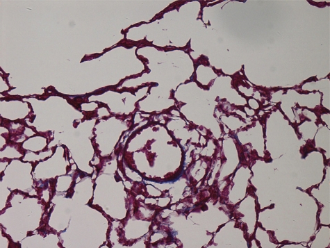
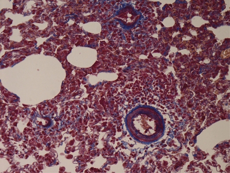
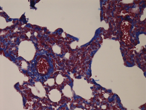
Results: Compared with control group, RVSP, percentage of fibrous tissue (F%) in pulmonary arterioles, mRNA levels of TGFß1, and receptors of CA-JVand MCT groups increased significantly. Severe hemodynamics change was found in MCT groups. On the other hand, CA-JV group demonstrated more obvious fibrogenesis and TGFß1 signals' upregulation in two pulmonary artery hypertension (PAH) models.
Conclusions: CA-JV shunt model of rats was a well-established PAH animal model simulating congenital heart disease with systemic-pulmonary shunt.
Figures
References
-
- Humbert M, Morrell NW, Archer SL, Stenmark KR, MacLean MR, Lang IM, Christman BW, Weir EK, Eickelberg O, Voelkel NF, Rabinovitch M. Cellular and molecular pathobiology of pulmonary arterial hypertension. J Am Coll Cardiol. 2004;43:13S–24S. - PubMed
-
- Lang IM, Klepetko W. Chronic thromboembolic pulmonary hypertension: an updated review. Curr Opin Cardiol. 2008;23:555–9. - PubMed
-
- Galie N, Manes A, Farahani KV, Pelino F, Palazzini M, Negro L, Romanazzi S, Branzi A. Pulmonary arterial hypertension associated to connective tissue diseases. Lupus. 2005;14:713–7. - PubMed
Publication types
MeSH terms
Substances
LinkOut - more resources
Full Text Sources
Medical
