. 2011 Dec;39(22):9789-802.
doi: 10.1093/nar/gkr639. Epub 2011 Sep 5.

Cation binding to 15-TBA quadruplex DNA is a multiple-pathway cation-dependent process

Affiliations

Cation binding to 15-TBA quadruplex DNA is a multiple-pathway cation-dependent process

Roman V Reshetnikov et al. Nucleic Acids Res. 2011 Dec.

Abstract

A combination of explicit solvent molecular dynamics simulation (30 simulations reaching 4 µs in total), hybrid quantum mechanics/molecular mechanics approach and isothermal titration calorimetry was used to investigate the atomistic picture of ion binding to 15-mer thrombin-binding quadruplex DNA (G-DNA) aptamer. Binding of ions to G-DNA is complex multiple pathway process, which is strongly affected by the type of the cation. The individual ion-binding events are substantially modulated by the connecting loops of the aptamer, which play several roles. They stabilize the molecule during time periods when the bound ions are not present, they modulate the route of the ion into the stem and they also stabilize the internal ions by closing the gates through which the ions enter the quadruplex. Using our extensive simulations, we for the first time observed full spontaneous exchange of internal cation between quadruplex molecule and bulk solvent at atomistic resolution. The simulation suggests that expulsion of the internally bound ion is correlated with initial binding of the incoming ion. The incoming ion then readily replaces the bound ion while minimizing any destabilization of the solute molecule during the exchange.


Figures

Figure 1.
Figure 1.
15-TBA fold. Two G-quartets, upper (G1, G6, G10 and G15) and lower (G2, G5, G11 and G14), form G-quadruplex. Lighter and darker tetragons show syn and anti stem nucleotides. The remaining nucleotides form one TGT and two TT loops. An ∼2-fold axis of symmetry relates the two halves of the G-quadruplex, resulting in two symmetric wide grooves (blue) and two symmetric narrow grooves (red).
Figure 2.
Figure 2.
Variants of 15-TBA-cation complexes used in QM/MM computations.
Figure 3.
Figure 3.
Sketch of ion penetration into the central binding site of 15-TBA through the top of the structure.
Figure 4.
Figure 4.
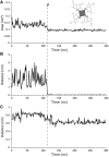
Structural dynamic of 15-TBA during the Na+ cation penetration through the upper part of the structure. (A) Area of a tetragon formed by O6 atoms of the upper G-quartet. Sketch of the tetragon (gray) is depicted over the graph. (B) Distance between the Na+ cation and center of mass (COM) of the eight O6 atoms of the G-quadruplex stem, i.e. the center of the quadruplex. (C) Distance between the COMs of the G8 nucleic acid base and the eight O6 atoms of the G-quadruplex stem. Similar graph for T9 is not shown. The letter ‘P’ and vertical dashed line mark the moment of the cation penetration into the quadruplex.
Figure 5.
Figure 5.
Penetration of potassium cation into substantially destabilized quadruplex.
Figure 6.
Figure 6.
Behavior of the TGT loop during potassium cation uptake from the bulk. Dotted line: distance between COMs of the K+ cation and the eight O6 atoms of the G-quadruplex stem. Solid black line: distance between COMs of the G8 nucleic base and the eight O6 atoms of the G-quadruplex. Solid gray line: distance between COMs of the T9 nucleobase and the eight O6 atoms of the G-quadruplex. Penetration of the cation into the G-stem (dotted line falls to 0 at 82 ns) correlates with G8 but not with T9 movement. The potassium cation did not spend any time in plane of the G-tetrad.
Figure 7.
Figure 7.
Complete exchange of sodium cation between the TT-free 15-TBA construct and bulk solution (snapshots are shown, the incoming and initially bound ions are shown as small black and gray dots, respectively).
Figure 8.
Figure 8.
Behavior of the ions in different complexes with 15-TBA in the QM/MM MD runs. Left: the distance between the cation and geometrical center of the G-quadruplex. The border of the central binding site is denoted by the magenta dotted line; if the distance is between 0 and the border, then the cation is inside the central binding site. Right: schematic presentation of cation movements. Cation colors correspond to the distance chart.
Figure 9.
Figure 9.
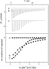
ITC curves of 15-TBA binding with potassium and barium. (A) Titration of 15-TBA by potassium after saturation with barium (competition), no effect; (B) binding of 15-TBA with potassium; (C) binding of 15-TBA with barium.

References

    1. Gellert M, Lipsett MN, Davies DR. Helix formation by guanylic acid. Proc. Natl Acad. Sci. USA. 1962;48:2013–2018. - PMC - PubMed
    1. Blackburn EH. Telomeres: no end in sight. Cell. 1994;77:621–623. - PubMed
    1. De Lange T. Telomere-related genome instability in cancer. Cold Spring Harb. Symp. Quant. Biol. 2005;70:197–204. - PubMed
    1. De Cian A, Lacroix L, Douarre C, Temime-Smaali N, Trentesaux C, Riou J-F, Mergny J-L. Targeting telomeres and telomerase. Biochimie. 2008;90:131–155. - PubMed
    1. Sen D, Gilbert W. Formation of parallel four-stranded complexes by guanine-rich motifs in DNA and its implications for meiosis. Nature. 1988;334:364–366. - PubMed

Publication types