. 2011 Sep 5;194(5):765-77.
doi: 10.1083/jcb.201011027.

Coatomer and dimeric ADP ribosylation factor 1 promote distinct steps in membrane scission

Affiliations

Coatomer and dimeric ADP ribosylation factor 1 promote distinct steps in membrane scission

Rainer Beck et al. J Cell Biol. .

Abstract

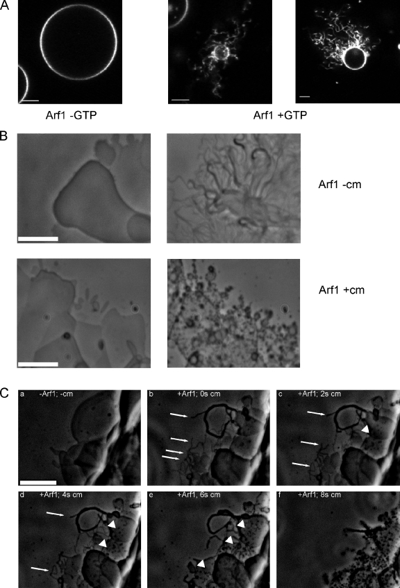
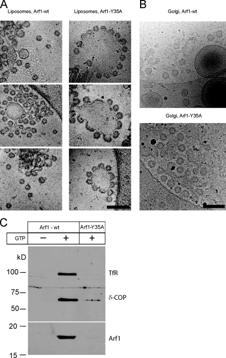
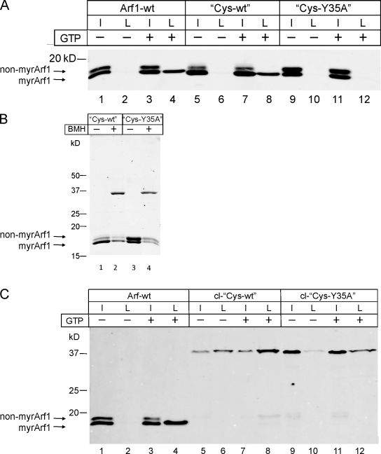
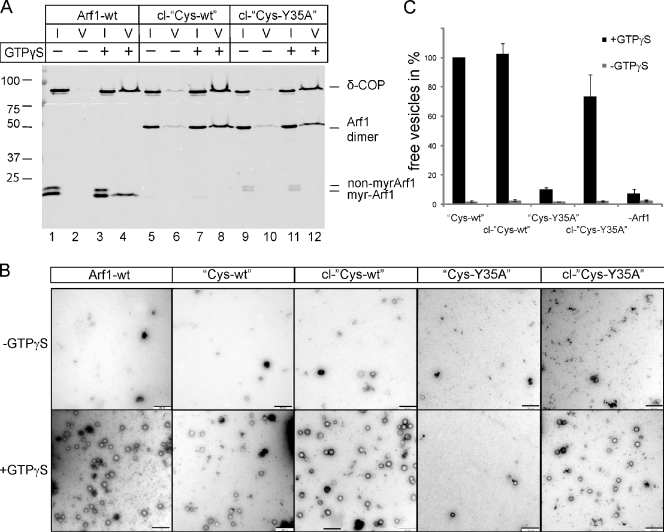
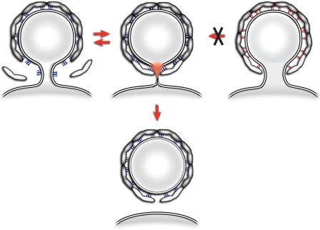
Formation of coated vesicles requires two striking manipulations of the lipid bilayer. First, membrane curvature is induced to drive bud formation. Second, a scission reaction at the bud neck releases the vesicle. Using a reconstituted system for COPI vesicle formation from purified components, we find that a dimerization-deficient Arf1 mutant, which does not display the ability to modulate membrane curvature in vitro or to drive formation of coated vesicles, is able to recruit coatomer to allow formation of COPI-coated buds but does not support scission. Chemical cross-linking of this Arf1 mutant restores vesicle release. These experiments show that initial curvature of the bud is defined primarily by coatomer, whereas the membrane curvature modulating activity of dimeric Arf1 is required for membrane scission.


Figures

Figure 1.
Figure 1.
Membrane surface activity of Arf1 and coatomer. (A) Membrane surface activity of Arf1 analyzed in GUVs. GUVs containing 0.5 mol% 1,2-dipalmitoyl-sn-glycero-3-phosphoethanolamine-N-lissamine rhodamine B sulfonyl were incubated with 3.5 µM Arf1-wt and 0.4 µM ARNO in the presence or absence of 1 mM GTP as indicated and recorded in a confocal laser-scanning microscope (LSM 510; Carl Zeiss) with a 63× objective lens and a pinhole size equivalent to one Airy disk diameter. (B) Membrane surface activity of Arf1 and coatomer. Lipids containing the p23 lipopeptide were spotted on a glass surface and hydrated with buffer containing GTP and 50 nM of the exchange factor ARNO. The lipid surface was observed before (left) and after addition of 1 µM myristoylated Arf1-GDP (top right) or by coinjection of 1 µM Arf1 and 0.25 µM coatomer (cm; bottom right). (C) Dissecting membrane surface activities of Arf1 and coatomer. (a) Lipids containing the p23 lipopeptide were spotted on a glass surface and hydrated with buffer containing GTP and the exchange factor ARNO as described in this legend. The image was taken, and thereafter, Arf1 was added, and tubule formation was observed (Video 1). (b) 0.25 µM coatomer was added to the chamber, leading to an immediate and nonspecific loss of most tubular structures caused by capillary flow forces. One frame afterward (t = 0), remaining Arf1-generated tubules are depicted (arrows). (c–f) Images were taken at 2, 4, 6, and 8 s after the addition of coatomer. The rapid degradation of Arf1-generated tubules (arrows) was followed over time, whereas new tubular structures with a distinct morphology were generated in the presence of coatomer (arrowheads). Bars, 5 µm.
Figure 2.
Figure 2.
COPI budding from synthetic liposomes and Golgi membranes analyzed by cryo-EM. (A and B) Cryo-EM of membranes incubated with Arf1-wt or Arf1-Y35A in the presence of the guanine nucleotide exchange factor ARNO, coatomer, and GTPγS. Images show liposomes (A) and Golgi membranes (B). Bars, 200 nm. (C) Purification of reconstituted of COPI vesicles. Recombinant Arf1-wt or Arf1-Y35A was incubated with Golgi membranes in the presence of GDP or GTP as in Fig. 2 B. Vesicles were purified by sucrose density centrifugation. Samples in the vesicle fraction were analyzed by SDS-PAGE and Western blotting using antibodies against the membrane marker transferrin receptor (TfR) and against coatomer subunit δ-COP and Arf1.
Figure 3.
Figure 3.
Binding of Arf1 and Arf1 variants to synthetic liposomes. (A) Comparison of the binding ability of Arf1-wt, Cys-wt, and Cys-Y35A to synthetic liposomes in the absence and presence of GTP. Arf1 variants were mixed with liposomes to a final concentration of 1.5 µM protein and 0.5 mM lipid in the presence of ARNO in a total volume of 100 µl. After 15 min of incubation at 37°C with or without 1 mM GTP, the samples were floated to an interface between 25 and 0% (wt/vol) sucrose. 10% of the collected liposomal fractions was compared with 5% of the input and analyzed for the presence of Arf1 and the Arf1 variants by SDS-PAGE and Western blotting, respectively. non-myrArf1, nonmyristoylated Arf1; myrArf1, myristoylated Arf1. (B) Cross-linking of the Cys variants using BMH. Purified Arf1 protein was mixed with BMH in a molar ratio of 2:1 and incubated for 1 h at RT. Thereafter, the cross-linking reaction was quenched by the addition of DTT and analyzed by SDS-PAGE and Coomassie staining. (C) Analysis of the binding ability of cross-linked Cys-wt (cl-Cys-wt) and Cys-Y35A (cl-Cys-Y35A) to synthetic liposomes in the absence and presence of GTP. The assay was performed as outlined in A. I, input; L, liposome-bound fraction.
Figure 4.
Figure 4.
Dimerization of Cys-Y35A restores its ability to generate COPI-coated vesicles from Golgi membranes. (A) COPI-coated vesicles were reconstituted from Golgi membranes using Arf1-wt, cross-linked Cys-wt (cl-Cys-wt), or cross-linked Cys-Y35A (cl-Cys-Y35A) in the presence of GTPγS and purified coatomer. Vesicles were purified via sucrose density centrifugation. 1% of input (I) and 50% of vesicle (V) fractions were analyzed by SDS-PAGE and Western blotting against δ-COP and Arf1. Non-myrArf1, nonmyristoylated Arf1; myr-Arf1, myristoylated Arf1. (B) Purified vesicles were analyzed by negative staining EM. Bars, 250 nm. (C) Quantification of vesicle formation: 14 image sections of 3-µm2 size were randomly chosen, and the number of vesicles was counted. Error bars represent the standard deviation of the mean of three independent experiments. Molecular masses are given in kilodaltons.
Figure 5.
Figure 5.
Interactions of Arf1 with coatomer probed by site-directed photo–cross-linking. Arf1 variants with a photolabile amino acid residue in either position 49 or 167 were prepared as described in Materials and methods and used for coatomer recruitment to Golgi membranes. Membranes were separated by centrifugation and UV irradiated followed by SDS-PAGE and Western blotting. (A) Analysis of photo–cross-link products with the photolabile amino acid derivative in position 49. Lanes 1 and 2 show Cys-wt and Cys-Y35A, respectively, decorated with anti-Arf1 antibodies. Lanes 3 and 4 show samples as in lanes 1 and 2 decorated with anti–γ-COP and, as a control, with anti–α-COP antibodies. (B) Analysis of photo–cross-link products with the photolabile amino acid derivative in position 167. Lanes 1 and 2 are decorated with anti-Arf1 antibodies, and lanes 3 and 4 are decorated with anti–δ-COP and anti–α-COP antibodies. For details of preparation of site-directed photolabile Arf1 derivatives see Materials and methods. Molecular masses are given in kilodaltons.
Figure 6.
Figure 6.
A model for Arf1-mediated membrane separation. The process of COPI budding and fission is depicted. Arf1 is recruited to the neck of a growing bud and stabilized in this location by coatomer (Arf1-Y35A in red; Arf1-wt in blue). As the growing bud becomes completed, the local membrane curvature creates an unfavorable situation for the insertion of the myristoylated amphipathic helix of Arf1. If Arf1 is enforced in this area by its multiple interactions with the covering network of polymerized coatomer, this will lead to local strain in the membrane (in red). The resulting metastable intermediate can then be relaxed in an irreversible manner by membrane separation. The resulting shell of polymerized coatomer is drawn with a gap because any possible contribution to membrane fission by closing this gap is not addressed here.

References

    1. Ambroggio E., Sorre B., Bassereau P., Goud B., Manneville J.B., Antonny B. 2010. ArfGAP1 generates an Arf1 gradient on continuous lipid membranes displaying flat and curved regions. EMBO J. 29:292–303 10.1038/emboj.2009.341 - DOI - PMC - PubMed
    1. Antonny B., Huber I., Paris S., Chabre M., Cassel D. 1997. Activation of ADP-ribosylation factor 1 GTPase-activating protein by phosphatidylcholine-derived diacylglycerols. J. Biol. Chem. 272:30848–30851 10.1074/jbc.272.49.30848 - DOI - PubMed
    1. Austin C., Hinners I., Tooze S.A. 2000. Direct and GTP-dependent interaction of ADP-ribosylation factor 1 with clathrin adaptor protein AP-1 on immature secretory granules. J. Biol. Chem. 275:21862–21869 10.1074/jbc.M908875199 - DOI - PubMed
    1. Austin C., Boehm M., Tooze S.A. 2002. Site-specific cross-linking reveals a differential direct interaction of class 1, 2, and 3 ADP-ribosylation factors with adaptor protein complexes 1 and 3. Biochemistry. 41:4669–4677 10.1021/bi016064j - DOI - PubMed
    1. Barlowe C., Orci L., Yeung T., Hosobuchi M., Hamamoto S., Salama N., Rexach M.F., Ravazzola M., Amherdt M., Schekman R. 1994. COPII: a membrane coat formed by Sec proteins that drive vesicle budding from the endoplasmic reticulum. Cell. 77:895–907 10.1016/0092-8674(94)90138-4 - DOI - PubMed

Publication types

MeSH terms