. 2012 May;90(5):540-52.
doi: 10.1038/icb.2011.71. Epub 2011 Sep 6.

ISCOMATRIX vaccines mediate CD8+ T-cell cross-priming by a MyD88-dependent signaling pathway

Affiliations
Free PMC article

ISCOMATRIX vaccines mediate CD8+ T-cell cross-priming by a MyD88-dependent signaling pathway

Nicholas S Wilson et al. Immunol Cell Biol. 2012 May.
Free PMC article

Abstract

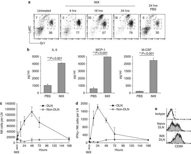
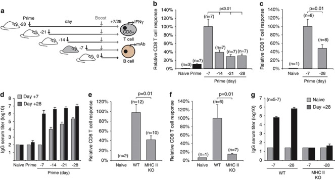
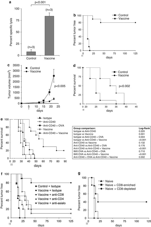
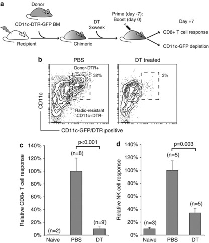
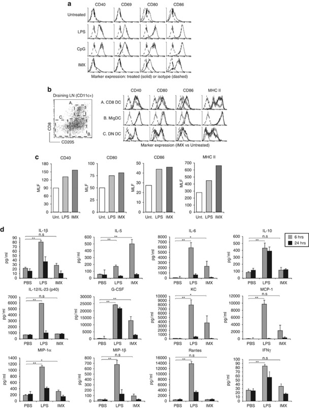
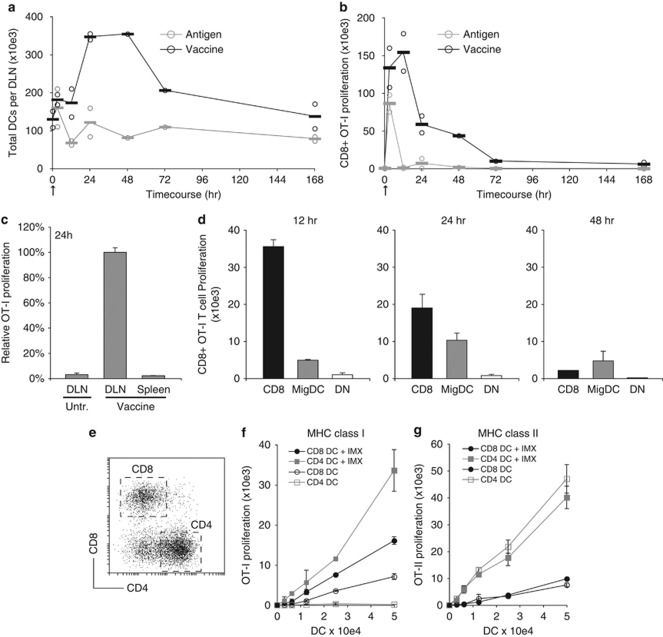
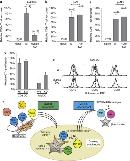
Generating a cytotoxic CD8(+) T-cell response that can eradicate malignant cells is the primary objective of cancer vaccine strategies. In this study we have characterized the innate and adaptive immune response to the ISCOMATRIX adjuvant, and the ability of vaccine antigens formulated with this adjuvant to promote antitumor immunity. ISCOMATRIX adjuvant led to a rapid innate immune cell response at the injection site, followed by the activation of natural killer and dendritic cells (DC) in regional draining lymph nodes. Strikingly, major histocompatibility complex (MHC) class I cross-presentation by CD8α(+) and CD8α(-) DCs was enhanced by up to 100-fold when antigen was formulated with ISCOMATRIX adjuvant. These coordinated features enabled efficient CD8(+) T-cell cross-priming, which exhibited prophylactic and therapeutic tumoricidal activity. The therapeutic efficacy of an ISCOMATRIX vaccine was further improved when co-administered with an anti-CD40 agonist antibody, suggesting that ISCOMATRIX-based vaccines may combine favorably with other immune modifiers in clinical development to treat cancer. Finally, we identified a requirement for the myeloid differentiation primary response gene 88 (MyD88) adapter protein for both innate and adaptive immune responses to ISCOMATRIX vaccines in vivo. Taken together, our findings support the utility of the ISCOMATRIX adjuvant for use in the development of novel vaccines, particularly those requiring strong CD8(+) T-cell immune responses, such as therapeutic cancer vaccines.


Figures

Figure 1
Figure 1
ISCOMATRIX adjuvant activates innate immune cells in vivo. (a) Immune cell infiltrates were gated on the myeloid marker CD11b. Monocyte (CD11b+Ly6C+) and neutrophil (CD11b+Gr1+) recruitment into subcutaneous air-pouches was evaluated 4, 16 and 24 h following subcutaneous ISCOMATRIX adjuvant (IMX) administration. Profiles are representative for n=3–6 mice per time-point. (b) Levels of IL-5, monocyte chemotactic protein-1 α and macrophage CSF (macrophage-CSF) detected in cultured air-pouch immune exudates. Error bars show the s.e.m. (n=3 per group). Student's t-test was used to calculate statistical significance. (c) Time-course of NK cell accumulation in the DLN (brachial) and non-DLN (inguinal) following subcutaneous IMX injection. (d) Ex vivo NK cell IFN-γ production in the DLN and non-DLN after IMX administration. Error bars represent the s.e.m. (n=4–8 individual LN per time-point). (e) Representative profiles showing NK cell expression of CD69 in the DLN of naïve or 24 h after IMX injection. No increase in CD69 expression was observed on NK cells, or other cell populations, from the non-DLN (data not shown). All results are representative of at least two independent experiments.
Figure 2
Figure 2
Cellular and humoral immune responses to ISCOMATRIX vaccines are dependent on CD4+ T-cell help. (a) Schematic showing the dosing regimen used to evaluate vaccine antigen-specific CD8+ T cell and antibody response. (b) IFN-γ production by endogenous OVA-specific CD8+ T cells was determined in the spleen 7 days after the boost vaccination. (c) IFN-γ production by endogenous gB (HSV-1)-specific CD8+ T cells was determined in the spleen 7 days after the boost vaccination. Results in (b, c) are expressed relative to the response obtained with a day −7, 0-dosing regimen. Mean values are expressed ±s.e.m.. Student's t-test was used to calculate statistical significance. (d) OVA-specific antibody titers (total IgG) in serum collected from mice vaccinated with the dosing protocols shown in (a). OVA-specific titers were quantified 7 or 28 days after the final vaccine dose. (e) OVA-specific CD8+ T-cell responses were compared in wild type (WT) and MHC class II-deficient mice (MHC II KO) vaccinated on days −7, 0. (f) The OVA-specific ‘recall' CD8+ T-cell response was assessed using a 5-day ex vivo re-stimulation protocol. (g) OVA-specific IgG titers in serum collected from naïve WT or MHC II KO vaccinated with two different vaccine dosing regimens. All results are representative of at least two or more independent experiments. ‘Student's' t-test was used to calculate statistical significance.
Figure 3
Figure 3
The antitumor response mediated by ISCOMATRIX vaccines requires components of innate and adaptive immunity. (a) The CTL response was evaluated in animals that received the ISCOMATRIX (IMX) vaccine or antigen (OVA) alone on day −7, 0. SIINFEKL-pulsed CFSEhigh-labeled target cells were injected intravenously on day +7, and specific-lysis was evaluated ex vivo 4 h later by flow cytometry. Specific lysis was calculated relative to a control (non-pulsed) CFSElow-labeled cell population. The mean specific lysis is shown ±s.e.m. Student's t-test was used to calculate statistical significance. (b) Tumor incidence in mice that received IMX vaccine (n=9) or IMX alone (control, n=10) on day −7, 0. All animals were challenged 7 days after boost dose with 1.5 × 10⁵ B16:OVA melanoma cells. (c) Tumor cells were inoculated in mice 5 days before receiving the IMX vaccine (vaccine) or IMX alone (control) on day 5 and 12. Tumor volumes were monitored until the first tumor reached the 3000 mm3, which was nominated as the survival end-point. ‘Student's' t-test was used to calculate statistical significance; n=10 per group. (d) Kaplan–Meier curves showing survival end-points for the cohorts in (c) (P<0.002 by the log-rank test) (n=10 per group). (e) Tumor cells were inoculated into mice 5 days before the indicated vaccination regimens (day 5 (prime) and 12 (boost)). Kaplan–Meier curves showing survival end-points for each cohort (n=10 per group; vaccine versus anti-CD40+vaccine=P<0.002 by the log-rank test). (f) Mice were administered anti-CD4, anti-CD8 or anti-asialo-GM1 (anti-asialo) on days −8, −5, −2, +1 and +4 during the day −7, 0 vaccine regimen. Mice were challenged on day +7 with 1.5 × 10⁵ B16:OVA cells and tumor incidence was monitored. (g) CD8-enriched or CD8-depleted spleen cell fractions from pre-vaccinated mice were adoptively transferred into naïve recipients. After 1 day of adoptive transfer, mice were challenged with 5 × 10⁵ B16:OVA melanoma cells. Tumor incidence was monitored out to day 125 (n=10–12 per group). All results are representative of two or more independent experiments.
Figure 4
Figure 4
CD11c-expressing cells are required for ISCOMATRIX adjuvant -mediated NK cell and CD8+ T-cell responses. (a) Schematic showing the generation of CD11c-DTR mice, and the CD11c+ depletion protocol used during the prime and boost IMX vaccine regimen. (b) Representative profile showing the depletion of radiosensitive CD11c/DTR-green fluorescent protein-expressing DCs at the time the antigen-specific CD8+ T-cell immune responses were quantified. (c) CD11c-DTR mice were either treated with PBS or DT 1 day before the prime vaccine dose, and then three times per week. The OVA-specific CD8+ T-cell response was assessed ex vivo in the spleen 7 days after the boost vaccination. The magnitude of the response is shown relative to the PBS-treated cohort ±s.e.m. Results are pooled from two separate experiments. (d) CD11c-DTR mice were treated with PBS or DT −3 and −1 days before a single dose of ISCOMATRIX adjuvant. Ex vivo NK cell IFN-γ production was measured in the DLN after 24 h. The magnitude of the response is shown relative to the PBS-treated cohort, ±s.e.m. The results are representative of at least two separate experiments. Student's t-test was used to calculate statistical significance.
Figure 5
Figure 5
In vivo DC activation and cytokine responses to ISCOMATRIX adjuvant administration. (a) Flt3L-derived DCs were cultured overnight in the presence of IMX (5 μg ml−1), CpG (1 μM) or LPS (1 μg ml−1). CD40, CD69, CD80 and CD86 upregulation (black lines) was monitored on conventional (CD11c+CD45RA) DCs by flow cytometry, as compared with an isotype control antibody (dashed lines). (b) CD8 and double-negative (DN) lymphoid DCs were distinguished from ‘tissue-derived' MigDCs based on the expression of CD8 and CD205 (left dot plot). Right histogram; expression of CD40, CD80, CD86 and MHC class II expression (black lines) on DCs following a single subcutaneous dose of ISCOMATRIX adjuvant, as compared with DCs isolated from untreated DLN (gray lines). Isotype control antibody staining is shown as dashed lines. Data are representative of 3–5 experiments. (c) Activation marker expression by CD8 DCs isolated from the DLN of untreated mice, or 24 h after a subcutaneous dose of IMX (5 μg) or LPS (3 μg). The mean linear fluorescence (MLF) is shown on the y-axis, with isotype controls MLF values subtracted for each sample. (d) Cytokine levels in the serum collected 6 or 24 h after subcutaneous IMX or LPS administration: shown are the levels of IL—1β,IL-5,IL-6, IL-10, IL-12/23(p40), granulocyte-CSF, keratinocyte chemoattractant (KC or CXCL1), monocyte chemotactic protein-1 (MCP-1 or CCL2), macrophage inflammatory protein-1α (MIP-1α or CCL3) and β (MIP-1β or CCL4), Rantes (or C) and IFN-γ. Error bars show the s.e.m. (n=6 mice per treatment group). Student's t-test was used to calculate statistical significance (*P<0.05, **P<0.001, n.s., not significant). All results are representative of two or more independent experiments.
Figure 6
Figure 6
ISCOMATRIX adjuvant facilitates antigen entry into the MHC class I cross-presentation pathway in DCs. (a) Time-course showing the number of CD11c+ DCs isolated from the DLN of mice injected with vaccine or OVA (antigen) alone. (b) MHC class I cross-presentation was quantified in the DLN ex vivo after a single dose of ISCOMATRIX vaccine or OVA (antigen) alone. The CD11c+ DC fraction (>90%) was purified from the DLN at the indicated times. A total of 5 × 10³ DCs were then co-cultured with 5 × 10⁴ CFSE-labeled OT-1 CD8+ T cells. Proliferation was quantified 60 h later by flow cytometry. Circles in (a) and (b) represent data points from independent experiments (an average of n=4–8 DLN per sample), and bars show the pooled average. (c) MHC-I presentation by CD11c+ DCs purified from the spleen or DLN 24 h after a single ISCOMATRIX vaccine dose. OT-I proliferation was determined as in (b). (d) Highly purified (>95%) populations of CD8, DN and MigDC were purified from the DLN 12, 24 and 48 h after a single vaccine dose. Cross-presentation was assessed by co-culturing each DC population with 5 × 10⁴ CFSE-labeled OT-1 cells and quantifying proliferation 60 h later. (e) CD11c-enriched spleen DCs were separated into CD8 and CD4 populations by flow cytometry (>95% purity). (f) CFSE-labeled OT-I T cells were co-cultured with CD8 or CD4 DCs pulsed for 30 min with OVA (antigen) alone (open symbols) or antigen+ISCOMATRIX (closed circles). Pulsed DCs were cultured with CFSE-labeled OT-I cells, and proliferation was determined after 60 h as described. (g) The same DCs used in (f) were co-cultured with CFSE-labeled CD4+ OT-II T cells. Proliferation was quantified as described. Error bars represent the s.d. of triplicate samples. Results are representative of two or more independent experiments.
Figure 7
Figure 7
ISCOMATRIX vaccines are dependent on a MyD88-signaling axis in vivo (a) CD8+ T-cell responses were compared in wild type (WT) and MyD88-deficient mice (MyD88 KO) vaccinated with an ISCOMATRIX vaccine on day −7, 0, with the magnitude of the CD8+ T-cell response shown relative to WT mice. (b, c) Same as in (a) except the CD8+ T-cell response was evaluated in TRIF or TLR4-deficient mice. (d) Purified CD8 and MigDCs from wild type or MyD88-deficient (KO) mice were isolated from the DLN 24 h after vaccine administration. MHC class I cross-presentation was assessed by co-culturing each population with 5 × 10⁴ CFSE-labeled OT-1 cells and quantifying proliferation 60 h later, as described. (e) CD40, CD80 and CD86 expression (black lines) was assessed for CD8 DCs isolated from the DLN of WT or MyD88 KO mice dosed with ISCOMARTIX adjuvant, compared with CD8 DCs from untreated WT mice (gray lines). Dashed lines illustrate the median fluorescence for each marker. (f) Schematic illustrating the interaction between DCs, T cells and NK cells in the DLN following ISCOMATRIX vaccine delivery. ISCOMATRIX vaccines initiate a localized inflammatory response at the subcutaneous injection site, and efficient DC activation and MHC class I cross-presentation in the DLN (MyD88-independent). Although the precise DC activation signal(s) is currently unknown, a distinct pro-inflammatory milieu was detected locally and systemically following ISCOMATIRX adjuvant administration. In the DLN, NK cell activation and CD8+ T-cell cross-priming was dependent on DCs, as well as an intact MyD88 signaling network. Cross-primed CD8+ T cells exhibit potent antitumor activity in prophylactic tumor challenge models. However, in the case of pre-established tumor burden, the effectiveness of the vaccine is likely to be blunted by immune suppressive networks, such as myeloid-derived suppressor cells (MDSC), regulatory T cells (Treg) and tumor-derived factors that prevent complete tumor eradication.

References

    1. Cheever MA, Allison JP, Ferris AS, Finn OJ, Hastings BM, Hecht TT, et al. The prioritization of cancer antigens: a national cancer institute pilot project for the acceleration of translational research. Clin Cancer Res. 2009;15:5323–5337. - PMC - PubMed
    1. Rosenberg SA, Yang JC, Restifo NP. Cancer immunotherapy: moving beyond current vaccines. Nat Med. 2004;10:909–915. - PMC - PubMed
    1. Lake RA, Robinson BW. Immunotherapy and chemotherapy—a practical partnership. Nat Rev Cancer. 2005;5:397–405. - PubMed
    1. Heath WR, Belz GT, Behrens GM, Smith CM, Forehan SP, Parish IA, et al. Cross-presentation, dendritic cell subsets, and the generation of immunity to cellular antigens. Immunol Rev. 2004;199:9–26. - PubMed
    1. Tritto E, Mosca F, De Gregorio E. Mechanism of action of licensed vaccine adjuvants. Vaccine. 2009;27:3331–3334. - PubMed

MeSH terms