

Pre-clinical evaluation of fluorescent deoxyglucose as a topical contrast agent for the detection of Barrett's-associated neoplasia during confocal imaging
- PMID: 21895028
- PMCID: PMC4527376
- DOI: 10.7785/tcrt.2012.500220
Pre-clinical evaluation of fluorescent deoxyglucose as a topical contrast agent for the detection of Barrett's-associated neoplasia during confocal imaging
Abstract
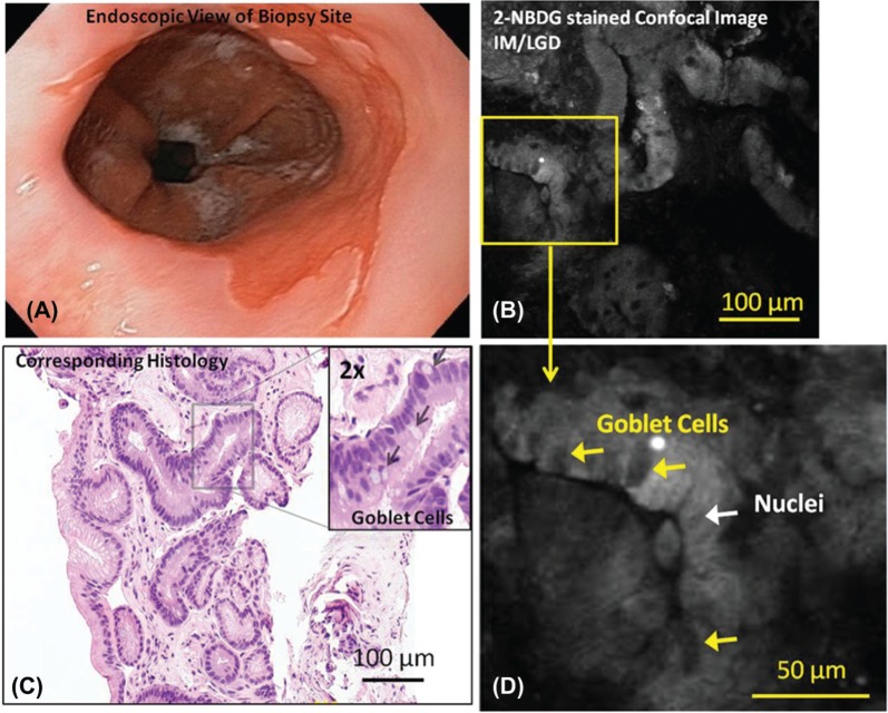
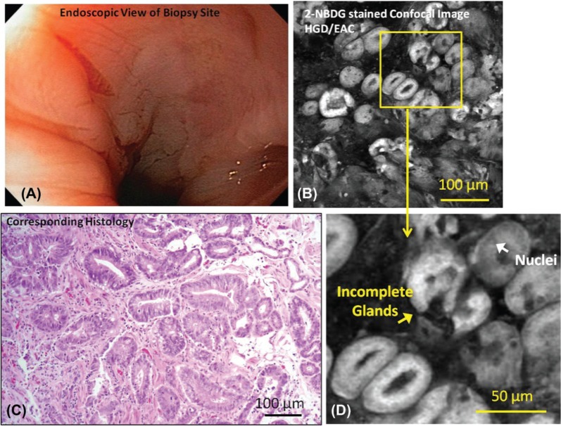
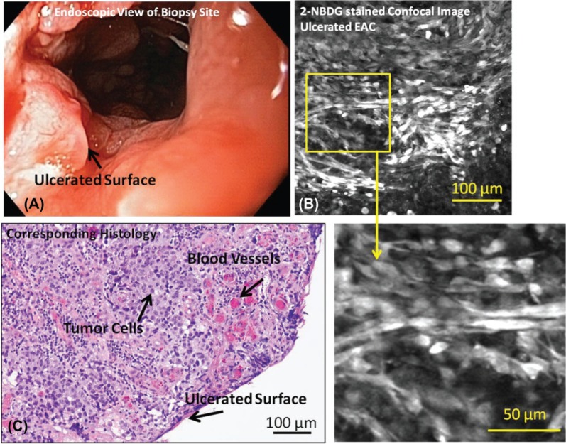
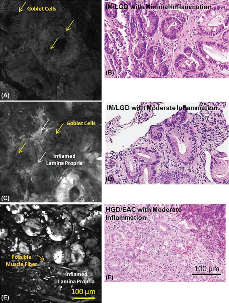
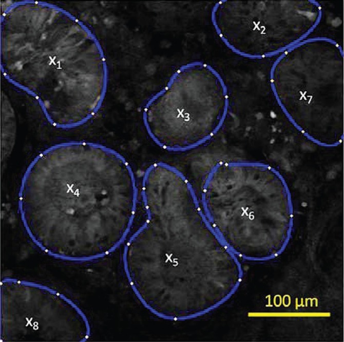
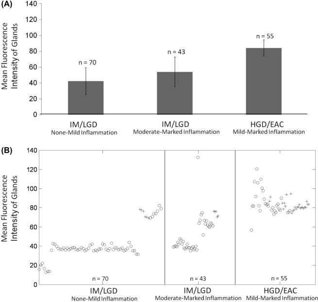
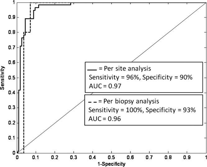
The availability of confocal endomicroscopy motivates the development of optical contrast agents that can delineate the morphologic and metabolic features of gastrointestinal neoplasia. This study evaluates 2-NBDG, a fluorescent deoxyglucose, the uptake of which is associated with increased metabolic activity, in the identification of Barrett's-associated neoplasia. Surveillance biopsies from patients with varying pathologic grades of Barrett's esophagus were incubated ex vivo at 37°C with 2-NBDG and imaged with a fluorescence confocal microscope. Images were categorized as neoplastic (high grade dysplasia, esophageal adenocarcinoma) or metaplastic (intestinal metaplasia, low grade dysplasia) based on the degree of glandular 2-NBDG uptake. Classification accuracy was assessed using histopathology as the gold standard. Forty-four biopsies were obtained from twenty-six patients; 206 sites were imaged. The glandular mean fluorescence intensity of neoplastic sites was significantly higher than that of metaplastic sites (p<0.001). Chronic inflammation was associated with increased 2-NBDG uptake in the lamina propria but not in glandular epithelium. Sites could be classified as neoplastic or not with 96% sensitivity and 90% specificity based on glandular mean fluorescence intensity. Classification accuracy was not affected by the presence of inflammation. By delineating the metabolic and morphologic features of neoplasia, 2-NBDG shows promise as a topical contrast agent for confocal imaging. Further in vivo testing is needed to determine its performance in identifying neoplasia during confocal endomicroscopic imaging.
Conflict of interest statement
Figures
References
-
- Cameron A. J. Epidemiology of Barrett’s esophagus and adenocarcinoma. Dis Esophagus 15, 106–108 (2002). - PubMed
-
- DeVault K. R. Epidemiology and significance of Barrett’s esophagus. Dig Dis 18, 195–202 (2000). - PubMed
-
- Sihvo E. I., Luostarinen M. E., Salo J. A. Fate of patients with adenocarcinoma of the esophagus and the esophagogastric junction: a population-based analysis. Am J Gastroenterol 99, 419–424 (2004). - PubMed
-
- Farrow D. C., Vaughan T. L. Determinants of survival following the diagnosis of esophageal adenocarcinoma (United States). Cancer Causes Control 7, 322–327 (1996). - PubMed
-
- Portale G., Hagen J. A., Peters J. H., Chan L. S., DeMeester S. R., Gandamihardja T. A., DeMeester T. R. Modern 5-year survival of resectable esophageal adenocarcinoma: single institution experience with 263 patients. J Am Coll Surg 202, 588–596; discussion 596–588 (2006). - PubMed
Publication types
MeSH terms
Substances
Grants and funding
LinkOut - more resources
Full Text Sources
Other Literature Sources
Medical
