

Cerebrovascular connexin expression: effects of traumatic brain injury
- PMID: 21895483
- PMCID: PMC3172862
- DOI: 10.1089/neu.2011.1900
Cerebrovascular connexin expression: effects of traumatic brain injury
Abstract
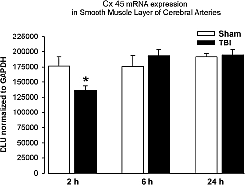
Traumatic brain injury (TBI) results in dysfunction of the cerebrovasculature. Gap junctions coordinate vasomotor responses and evidence suggests that they are involved in cerebrovascular dysfunction after TBI. Gap junctions are comprised of connexin proteins (Cxs), of which Cx37, Cx40, Cx43, and Cx45 are expressed in vascular tissue. This study tests the hypothesis that TBI alters Cx mRNA and protein expression in cerebral vascular smooth muscle and endothelial cells. Anesthetized (1.5% isoflurane) male Sprague-Dawley rats received sham or fluid-percussion TBI. Two, 6, and 24 h after, cerebral arteries were harvested, fresh-frozen for RNA isolation, or homogenized for Western blot analysis. Cerebral vascular endothelial and smooth muscle cells were selected from frozen sections using laser capture microdissection. RNA was quantified by ribonuclease protection assay. The mRNA for all four Cx genes showed greater expression in the smooth muscle layer compared to the endothelial layer. Smooth muscle Cx43 mRNA expression was reduced 2 h and endothelial Cx45 mRNA expression was reduced 24 h after injury. Western blot analysis revealed that Cx40 protein expression increased, while Cx45 protein expression decreased 24 h after injury. These studies revealed significant changes in the mRNA and protein expression of specific vascular Cxs after TBI. This is the first demonstration of cell type-related differential expression of Cx mRNA in cerebral arteries, and is a first step in evaluating the effects of TBI on gap junction communication in the cerebrovasculature.
Figures


References
-
- Armstead W.M. Kiessling J.W. Cines D.B. Higazi A.A. Glucagon protects against impaired NMDA-mediated cerebrovasodilation and cerebral autoregulation during hypotension after brain injury by activating cAMP protein kinase A and inhibiting upregulation of tPA. J. Neurotrauma. 2011;28:451–457. - PMC - PubMed
-
- Armstead W.M. Role of endothelin-1 in age-dependent cerebrovascular hypotensive responses after brain injury. Am. J. Physiol. 1999;274:H1884–H1894. - PubMed
-
- Bazan N.G. Rodriguez de Turco E.B. Allan G. Mediators of injury In Neurotrauma: intracellular signal transduction, gene expression. J. Neurotrauma. 1995;12:791–814. - PubMed
-
- Blanc E.M. Bruce-Keller A.J. Mattson M.P. Astrocytic gap junctional communication decreases neuronal vulnerability to oxidative stress-induced disruption of Ca2+ homeostasis and cell death. J. Neurochem. 1998;70:958–970. - PubMed
-
- Bouma G.J. Muizelaar J.P. Relationship between cardiac output and cerebral blood flow in patients with intact and with impaired autoregulation. J. Neurosurg. 1990;73:368–374. - PubMed
Publication types
MeSH terms
Substances
Grants and funding
LinkOut - more resources
Full Text Sources
Miscellaneous
