. 2011 Dec;17(12):1223-32.
doi: 10.1089/ten.tec.2011.0134. Epub 2011 Sep 6.

Three-dimensional system for the in vitro study of megakaryocytes and functional platelet production using silk-based vascular tubes

Affiliations

Three-dimensional system for the in vitro study of megakaryocytes and functional platelet production using silk-based vascular tubes

Isabella Pallotta et al. Tissue Eng Part C Methods. 2011 Dec.

Abstract

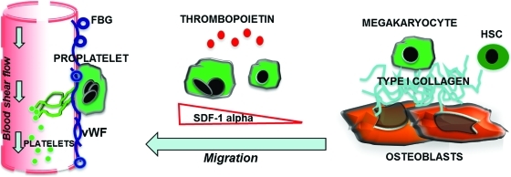
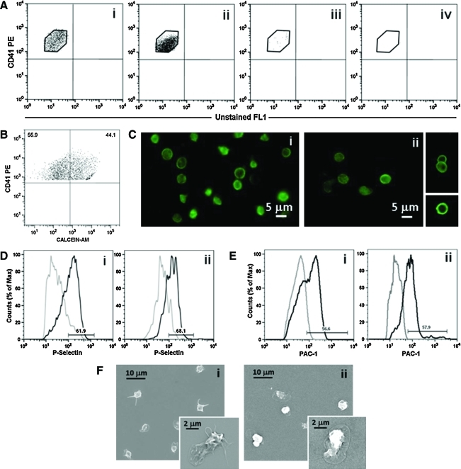
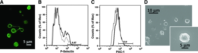
Platelets are specialized cells produced by megakaryocytes in the bone marrow that represent the first defense against hemorrhage, yet they also play a pathological role in thrombosis, inflammation, and cancer. Millions of platelet transfusions are conducted each year, and the supply of this blood component is limited. There are many diseases where platelet production or function is impaired with severe consequences for patients. With such clinical need, new insight into the formation of platelets would have a major impact on patients and healthcare. We developed an innovative 3D system to study platelet production that represents the first spatial reconstruction of the bone marrow environment. In this system human megakaryocytes were able to migrate toward the vascular niche, extend proplatelets, and release functional platelets into vascular tubes. The combination of different bone marrow components and the compliance of silk-based vascular tubes makes this model a unique tool for the study of platelet formation and production for use in healthcare needs.


Figures

FIG. 1.
FIG. 1.
Outline of platelet formation in the bone marrow environment. Immature megakaryocytes in contact with the osteoblastic niche are inhibited in their maturation. Upon migration toward the vascular niche, megakaryocytes extend proplatelets and release platelets into the blood stream. Interactions of megakaryocytes with matrices supposed to fill the vascular niche, such as fibrinogen (FBG) or von Willebrand factor (vWF), are able to sustain proplatelet formation, whereas type I collagen, in the osteoblastic niche, totally suppresses this event and prevents premature platelet release. SDF-1α is produced locally by the stromal cells and promotes the migration and contact of megakaryocytes with the permissive vascular niche. Color images available online at www.liebertonline.com/tec
FIG. 2.
FIG. 2.
Bioreactor platform and silk microtubes. (A) The previously developed bioreactor platform was adapted to reproduce the bone marrow microenvironment to study megakaryocyte migration, adhesion to the sinusoidal vessel, proplatelet formation, and platelet release. (B) Scanning electron microscopy (SEM) image of a representative silk microtube used to mimic blood vessels (ii). The wall thickness was adjusted around 50 μm to match proplatelet length (i). (C) To allow proplatelet elongation through the vascular microtube wall, defined pore sizes of 2–8 μm were obtained as described in Materials and Methods. (i) SEM image of vascular microtube wall pores. (ii) SEM image showing pore interconnection and a path through the tube wall. Color images available online at www.liebertonline.com/tec
FIG. 3.
FIG. 3.
Megakaryocyte migration, adhesion, and proplatelet formation. Mature megakaryocytes were stained with an anti CD61 antibody and added to each well of the bioreactor. (A) The silk microtubes were coated with 300 ng/mL SDF-1α and a combination of Matrigel with FBG and vWF or Matrigel alone or type I collagen, as controls. Prestained megakaryocytes were then added to each bioreactor and megakaryocyte migration toward the vascular microtubes was analyzed. After 16 h megakaryocyte migration through the gel composed of Matrigel+FBG+vWF was significantly higher compared with other conditions. (B) Z-stack projection (i) of megakaryocytes in adhesion to the surface of the silk microtube well after 24 h incubation with orthogonal projections in the z–y (ii) and x–z (iii) directions to confirm cell adhesion in tube cross-sections. (C) After an additional 16 h, megakaryocytes extended proplatelets through the vascular microtube wall as shown in the orthogonal projections of a proplatelet (green) protruding into the vascular tube (gray). Color images available online at www.liebertonline.com/tec
FIG. 4.
FIG. 4.
Released platelets inside silk microtubes are dependent on tube porosity. After staining megakaryocytes with an anti-CD61 antibody green and counterstaining nuclei with Hoechst 33258, cell suspension was added to bioreactors spanned with a porous (A) or nonporous (B) silk microtube coated with SDF-1α, Matrigel, FBG, and vWF. After 40 h incubation, released platelets were observed in immunofluorescence (Ai, Bi) by confocal analysis only inside porous tubes. As cells were counterstained with Hoechst 33258, we were able to demonstrate that large, nucleated megakaryocytes were not present inside the vascular microtube. Bright field of nonporous (Aii) and porous (Bii) silk microtubes show the tube edges. Yellow dotted lines indicate the tube edges. Color images available online at www.liebertonline.com/tec
FIG. 5.
FIG. 5.
Analysis of collected platelets. (A) Flow cytometry analysis of collected platelets in the microtube flow effluent: (ii) microtubes coated with SDF-1α and a combination of Matrigel, FBG, and vWF, (iii) uncoated microtubes, and (iv) microtubes coated with SDF-1α and type I collagen. Collected platelets were analyzed as CD41+ events with the same physical parameters and fluorescence intensity of human blood platelets (i). (B) To exclude fragments, collected cells were double-stained with an anti-CD41 antibody and calcein-AM. A representative dot plot is shown. (C) Circumferential microtubule coils were observed with an anti-tubulin antibody in the majority of collected platelets (ii) when compared to human peripheral blood platelets (i). (D, E) Platelets collected after bioreactor perfusion (Dii, Eii) revealed increased exposure of P-selectin to the plasma membrane and increased PAC-1 binding after thrombin stimulation, as in human peripheral blood platelets (Di, Ei) (gray line indicates unstimulated platelets; black line, thrombin-stimulated platelets). (F) Spreading of collected platelets after perfusion (ii) was analyzed by SEM in adhesion to type I collagen and compared to human peripheral blood platelets (i). Color images available online at www.liebertonline.com/tec
FIG. 6.
FIG. 6.
Analysis of in vitro released platelets in 2D culture. (A) Morphological analysis of in vitro released platelets. Immunofluorescence analysis by staining with an anti-tubulin antibody revealed the presence of dumbbells that were still dividing in culture. (B, C) Flow cytometry analysis of in vitro released platelet functionality. After activation with thrombin, in vitro released platelets showed low P-selectin expression (B) and PAC-1 binding (C) (gray line indicates unstimulated platelets; black line, thrombin-stimulated platelets). (D) SEM analysis revealed that the majority of in vitro released platelets were not able to spread in adhesion to type I collagen. Color images available online at www.liebertonline.com/tec

References

    1. AuBuchon J.P. Herschel L. Roger J. Further evaluation of a new standard of efficacy for stored platelets. Transfusion. 2005;45:1143. - PubMed
    1. Sullivan M.T. Cotton R. Read E.J. Wallace E.L. Blood collection and transfusion in the United States in 2001. Transfusion. 2007;47:385. - PubMed
    1. Avecilla S.T. Hattori K. Heissig B. Tejada R. Liao F., et al. Chemokine-mediated interaction of hematopoietic progenitors with the bone marrow vascular niche is required for thrombopoiesis. Nature Med. 2004;10:64. - PubMed
    1. Italiano J.E., Jr. Lecine P. Shivdasani R.A. Hartwig J.H. Blood platelets are assembled principally at the ends of proplatelet processes produced by differentiated megakaryocytes. J Cell Biol. 1999;147:1299. - PMC - PubMed
    1. Becker R.P. De Bruyn P.P. The transmural passage of blood cells into myeloid sinusoids and the entry of platelets into the sinusoidal circulation; a scanning electron microscopic investigation. Am J Anat. 1976;145:183. - PubMed

Publication types

LinkOut - more resources