. 2012 Mar 20;29(5):776-88.
doi: 10.1089/neu.2010.1602. Epub 2011 Nov 4.

Mitochondrial injury after mechanical stretch of cortical neurons in vitro: biomarkers of apoptosis and selective peroxidation of anionic phospholipids

Affiliations

Mitochondrial injury after mechanical stretch of cortical neurons in vitro: biomarkers of apoptosis and selective peroxidation of anionic phospholipids

Jing Ji et al. J Neurotrauma. .

Abstract

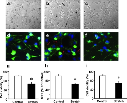
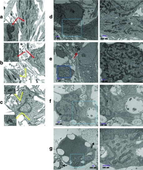
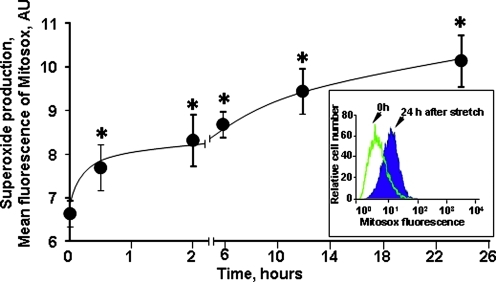
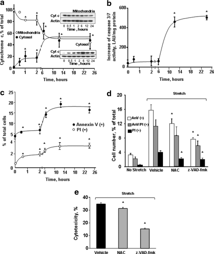
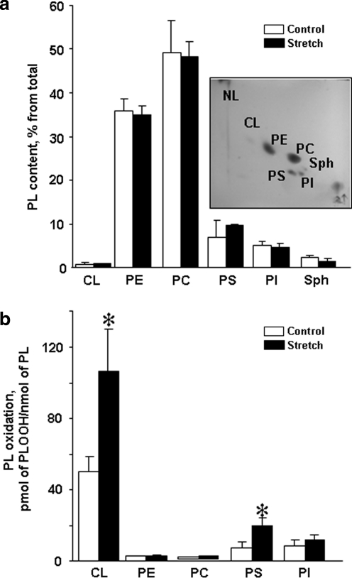
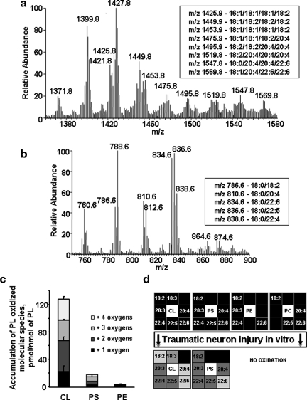
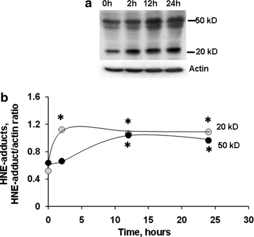
Mechanical injury of neurites accompanied by rupture of mitochondrial membranes may lead to immediate nonspecific release and spreading of pro-apoptotic factors and activation of proteases, that is, execution of apoptotic program. In the current work, we studied the time course of the major biomarkers of apoptosis as they are induced by exposure of rat cortical neurons to mechanical stretch. By using transmission electron microscopy, we found that mitochondria in the neurites were damaged early (1 h) after mechanical stretch injury whereas somal mitochondria were significantly more resistant and demonstrated structural damage and degenerative mitochondrial changes at a later time point after stretch (12 h). We also report that the stretch injury caused immediate activation of reactive oxygen species production followed by selective oxidation of a mitochondria-specific phospholipid, cardiolipin, whose individual peroxidized molecular species have been identified and quantified by electrospray ionization mass spectrometry analysis. Most abundant neuronal phospholipids - phosphatidylcholine, phophatidylethanolamine - did not undergo oxidative modification. Simultaneously, a small-scale release of cytochrome c was observed. Notably, caspase activation and phosphatidylserine externalization - two irreversible apoptotic events designating a point of no return - are substantially delayed and do not occur until 6-12 h after the initial impact. The early onset of reactive oxygen species production and cytochrome c release may be relevant to direct stretch-induced damage to mitochondria. The delayed emergence of apoptotic neuronal death after the immediate mechanical damage to mitochondria suggests a possible window of opportunity for targeted therapies.


Figures

FIG. 1.
FIG. 1.
Viability of cortical neurons after stretch injury. Representative photomicrographs of control cortical neurons (a, d) and cortical neurons after mechanical stretch, 10 s−1 strain rate and 50% membrane deformation (b, c, e, f). Stretch injury resulted in damage to the neuronal processes whereas the cell bodies remained intact and attached to the substrate, immediately after stretch (arrows, b, e), and at 24 h after stretch (c, f). Green: microtubule associated protein 2 (MAP2) immunostain; Blue: DAPI stain. Quantification of cell viability at 24 h after stretch injury using Alamar Blue (g) and MTT (h) assays and trypan blue (i) staining. Data are mean±SD, *p<0.05 vs control, n=6. Color image is available online at www.liebertonline.com/neu
FIG. 2.
FIG. 2.
Ultrastructural changes induced by mechanical stretch through transmission electron microscopy (TEM). Typical TEM image of control neurons (a) and cortical neurons 1h (b) and 12 h (c) after mechanical stretch. Representative TEM images are presented. Red (a, b) and yellow (b, c) arrows indicate normal and damaged mitochondria, respectively. (d) A representative section of control primary cortical neurons with dispersed chromatin and a clearly defined nucleolus. (e–g) A representative section of mechanically stretched primary cortical neurons showing apoptosis with intact plasma membrane, fragmented nuclei, chromatin clumping, and cytoplasmic vacuoles in 6 h (e), 12 h (f) and 24 h (g). Enlargement of the rectangular selected areas show the normal (d) or abnormal (f, g) mitochondria after mechanical stretch. Red arrow (e) indicates apoptotic bodies. Black arrows (g) indicate necrotic neurons with loss of membrane integrity and cell swelling. Color image is available online at www.liebertonline.com/neu
FIG. 3.
FIG. 3.
Time course of mitochondrial superoxide generation in cortical neurons after stretch injury assessed by flow cytometry. Insert: histograms demonstrating MitoSOX flouorescence intensities in control neurons and neurons 24 h after stretch. Data are mean±SEM, *p<0.005 vs. control, n=6. Color image is available online at www.liebertonline.com/neu
FIG. 4.
FIG. 4.
Mechanical stretch-induced apoptosis in cortical neurons. (a) Release of cyt c after stretch injury in cortical neurons. Typical Western blots (insets) and the densitometric analysis of cyt c in mitochondrial (closed circles) and cytosolic (open circles) fractions of neuronal cells. Data are mean±SD, *p<0.05 vs control, n=3. Note that release of cyt c from mitochondria occurred as early as 30 min after the the insult and peaked at 6 hrs after stretch injury. (b) Changes of caspase 3/7 activity in cortical neurons after stretch injury. Caspase 3/7 activity increased significantly at 12 h after stretch. The activity remained high at 24 h after injury. Data are mean±SD, *p<0.05 vs control, n=6. (c) Flow cytometry analysis of Annexin V and propidium iodide (PI) responses of cortical neurons to stretch. Early Annexin V-positivity (phosphatidylserine [PS] externalization) - indicating apoptosis – was markedly enhanced 6 h after stretch. PI positivity – corresponding to necrosis – followed the same trend with a lower magnitude. Data are mean±SD, *p<0.05 vs control, n=3. Effect of pre-treatment with antioxidant N-acetyl cysteine (NAC, 250 μM) and caspase inhibitor (z-VAD-fmk, 25 μM) on stretch-induced neuronal death assessed by flow cytometry analysis of PS externalization and PI positivity (d) and cytotoxicity (lactate dehydrogenase [LDH] release) relative to Triton exposure (e) at 24 h after stretch. Data are mean±SD, *p<0.05 vs respective vehicle group, n=4.
FIG. 5.
FIG. 5.
Phospholipid composition and accumulation of phospholipid hydroperoxides in rat cortical neurons after stretch. (a) Phospholipid composition of control and stretched neurons. Insert: Typical two-dimensional high performance thin layer chromatography (2D-HPTLC) of lipids extracted from rat cortical neurons. Total lipids were extracted and separated by 2D-HPTLC. (b) Content of phospholipid hydroperoxides in control and stretched neurons. Two hours after stretch, total lipids were extracted, separated by 2D-HPTLC and phospholipid hydroperoxides (PL-OOH) were determined using Amplex® Red protocol. *p<0.05 stretched vs. control neurons. PL, phospholipids; CL, cardiolipin; PE, phosphatidylethanolamine; PC, phosphatidylcholine; PS, phosphatidylserine; PI, phosphatidylinositol; Sph, Sphingomyelin.
FIG. 6.
FIG. 6.
Identification of phospholipid molecular species by liquid chromatography mass spectrometry (LC-MS). (a) Typical liquid chromatography electrospray ionization mass spectrometry (LC/ESI-MS) spectrum of CL isolated from control neurons. Insert: Major molecular species of CL containing polyunsaturated fatty acids. (b) Typical LC/ESI-MS spectrum of PS isolated from control neurons. Insert: Major molecular species of PS containing polyunsaturated fatty acids. (c) Accumulation of oxygenated phospholipids in rat cortical neurons 2 h after stretch assessed by LC-MS. Data are mean±SD, n=3. CL, cardiolipin; PS, phosphatidylserine; PE, phosphatidylethanolamine. (d) Oxidative lipidomics “Hit-map” of stretched neurons.
FIG. 7.
FIG. 7.
Formation of HNE-protein adducts in P2 fraction isolated from primary neurons after stretch injury. Using Western blot (a), anti-HNE antibody detected several protein bands with masses ranging from over 50 kD to less than 20 kD in cortical neurons cultured on elastic membranes. Increased levels of HNE protein adducts were observed after stretch (b). Data are mean±SD, *p<0.05 vs. respective control, n=3.

References

    1. Ainslie K.M. Tao S.L. Popat K.C. Desai T.A. In vitro immunogenicity of silicon-based micro- and nanostructured surfaces. ACS Nano. 2008;2:1076–1084. - PubMed
    1. Arundine M. Aarts M. Lau A. Tymianski M. Vulnerability of central neurons to secondary insults after in vitro mechanical stretch. J. Neurosci. 2004;24:8106–8123. - PMC - PubMed
    1. Barbee K.A. Macarak E.J. Thibault L.E. Strain measurements in cultured vascular smooth muscle cells subjected to mechanical deformation. Ann. Biomed. Eng. 1994;22:14–22. - PubMed
    1. Bayir H. Kapralov A.A. Jiang J. Huang Z. Tyurina Y.Y. Tyurin V.A. Zhao Q. Belikova N.A. Vlasova II. Maeda A. Zhu J. Na H.M. Mastroberardino P.G. Sparvero L.J. Amoscato A.A. Chu C.T. Greenamyre J.T. Kagan V.E. Peroxidase mechanism of lipid-dependent cross-linking of synuclein with cytochrome C: protection against apoptosis versus delayed oxidative stress in Parkinson disease. J. Biol. Chem. 2009;284:15,951–15,969. - PMC - PubMed
    1. Bayir H. Tyurin V.A. Tyurina Y.Y. Viner R. Ritov V. Amoscato A.A. Zhao Q. Zhang X.J. Janesko–Feldman K.L. Alexander H. Basova L.V. Clark R.S. Kochanek P.M. Kagan V.E. Selective early cardiolipin peroxidation after traumatic brain injury: an oxidative lipidomics analysis. Ann. Neurol. 2007;62:154–169. - PubMed

Publication types

MeSH terms

LinkOut - more resources