. 2012 Jan 15;16(2):139-52.
doi: 10.1089/ars.2011.3967. Epub 2011 Sep 22.

Chronic exercise preserves renal structure and hemodynamics in spontaneously hypertensive rats

Affiliations

Chronic exercise preserves renal structure and hemodynamics in spontaneously hypertensive rats

Deepmala Agarwal et al. Antioxid Redox Signal. .

Abstract

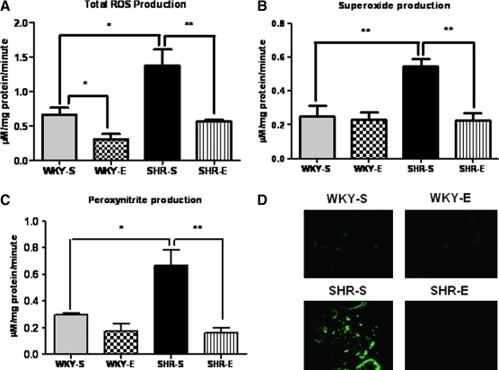
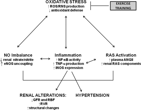
Aims: Exercise training (ExT) is a recommended adjunct to many pharmaceutical antihypertensive therapies. The effects of chronic ExT on the development of hypertension-induced renal injury remain unknown. We examined whether ExT would preserve renal hemodynamics and structure in the spontaneously hypertensive rat (SHR), and whether these effects were mediated by improved redox status and decreased inflammation. Normotensive WKY rats and SHR underwent moderate-intensity ExT for 16 weeks. One group of SHR animals was treated with hydralazine to investigate the pressure-dependent/independent effects of ExT. Acute renal clearance experiments were performed prior to sacrifice. Tissue free radical production rates were measured by electron paramagnetic resonance; gene and protein expression were measured by real time RT-PCR and Western blot or immunofluorescence, respectively. Plasma angiotensin II levels and kidney antioxidants were assessed. Training efficacy was assessed by citrate synthase activity assay in hind-limb muscle.

Results: ExT delayed hypertension, prevented oxidative stress and inflammation, preserved antioxidant status, prevented an increase in circulating AngII levels, and preserved renal hemodynamics and structure in SHR. In addition, exercise-induced effects, at least, in part, were found to be pressure-independent.

Innovation: This study is the first to provide mechanistic evidence for the renoprotective benefits of ExT in a model of hypertension. Our results demonstrate that initiation of ExT in susceptible patients can delay the development of hypertension and provide renoprotection at the functional and ultrastructural level.

Conclusion: Chronic ExT preserves renal hemodynamics and structure in SHR; these effects are partially mediated by improved redox status and decreased inflammation.


Figures

FIG.1.
FIG.1.
Citrate synthase activity (nmol/min/mg of protein) in soleus muscle of sedentary or exercised SHR and WKY as measured by citrate synthase activity assay kit (n=8 per group). After the period of 16 weeks of exercise, the activity of citrate synthase in the soleus muscle was significantly higher in SHR, as well as in WKY rats compared with their sedentary control groups, indicating the efficacy of the exercise protocol. ***p<0.001.
FIG. 2.
FIG. 2.
Assessment of glomerular injury, and urinary albumin and creatinine levels. (A–E) Representative photomicrographs for scoring of glomerular injury; scale bars=50 μm. (A) Grade 0: glomerulus with no lesions; (B) Grade 1: 0%–25% of glomerular area affected; (C) Grade 2: 25%–50% of glomerular area affected; (D) Grade 3: 50%–75% of glomerular area affected; (E) Grade 4: 75%–100% of glomerular area affected. (F) Average glomerular lesion scores for each experimental group (n=5–6 per group). SHR-S rats had more glomeruli with greater degrees of injury; these changes were prevented with ExT. (G, H) Urinary albumin and creatinine levels. SHR-S in comparison with WKY-S had significantly increased levels of albumin and creatinine in urine, indicating impairment in renal function, whereas SHR-E had significantly reduced levels when compared to SHR-S. $p<0.05 vs. WKY-S; *p<0.05 vs. SHR-S; p<0.05 vs. SHR-E. (To see this illustration in color the reader is referred to the web version of this article at www.liebertonline.com/ars).
FIG. 3.
FIG. 3.
Effects of ExT on renal tissue architecture and fibrosis. (A) Assessment of kidney tissue architecture, and interstitial and perivascular fibrosis by Masson's trichrome staining (n=5–6 per group), scale bars=200 μm. In comparison to WKY-S, SHR-S have more arterial myointimal hyperplasia (arrow), more tubular degeneration and ectasia (arrowhead), more interstitial, periglomerular, and periarterial fibrosis (light blue staining areas), and more glomerular parietal metaplasia (asterisk). Whereas, when compared to SHR-S, SHR-E rats exhibited less arterial myointimal hyperplasia, less tubular degeneration and ectasia, less interstitial, periglomerular, and periarterial fibrosis, and less glomerular parietal metaplasia. In WKY, there were no appreciable differences between WKY-S and WKY-E in aforementioned renal structural features except presence of less tubular degeneration and ectasia in WKY-E in comparison with WKY-S. (B) A representative Western blot and densitometric analyses of protein expression of collagen I in renal cortex. Our results revealed increased protein levels of collagen I in SHR-S in comparison with WKY-S, and ExT resulted in significant reduction in its level in SHRs. **p<0.05; ***p<0.001. (To see this illustration in color the reader is referred to the web version of this article at www.liebertonline.com/ars).
FIG. 4.
FIG. 4.
ExT attenuates production of oxidants in the kidney of SHRs. (A) Total reactive oxygen species (ROS), (B) superoxide, and (C) peroxynitrite production rates in renal cortical tissues of rats from each experimental group (n=6 per group), as determined by electron paramagnetic resonance spectroscopy. Chronic ExT prevented the increases in total ROS, superoxide, and peroxynitrite production rates normally seen in SHR. (D) Immunofluorescence (n=5–6 per group) for 3-nitrotyrosine, an indirect indicator of peroxynitrite formation. Exercise intervention prevented the increases in 3-NT normally seen in SHR. *p<0.01; **p<0.05. (To see this illustration in color the reader is referred to the web version of this article at www.liebertonline.com/ars).
FIG. 5.
FIG. 5.
ExT improves antioxidant levels in the kidney of SHRs. Levels of (A) reduced glutathione (GSH), (B) oxidized glutathione (GSSG), (C) GSH/GSSG ratio, and (D) glutathione peroxidase (GPx) as measured in cortical tissues from 8 rats from each experimental group. GSH, GSSG, and GPx levels were significantly reduced in SHR-S animals; levels were normalized in SHR-E animals. *p<0.01; **p<0 .05; ***p<0.001.
FIG. 6.
FIG. 6.
Protein levels of Cu/Zn-SOD and heme-oxygenase-1 (HO-1) in renal cortical tissue from each group (n=6 per group) as measured by Western blot analyses. There was no significant difference in the basal levels of renal Cu/Zn-SOD expression between WKY-S and SHR-S, however, WKY-E had significantly increased Cu/Zn-SOD levels compared with WKY-S. HO-1 levels were not significantly different among all four groups; however, SHR animals exhibited slightly higher levels compared to WKY rats.*p<0.01.
FIG. 7.
FIG. 7.
Effects of ExT on NF-κB p65 DNA binding activity and TNF-α levels in the kidney. (A) DNA binding activity of NF-κB p65 in renal cortical tissues of rats from each experimental group (n=6–8 per group), as determined by ELISA. $p<0.05 vs. WKY-S; *p<0.05 vs. SHR-S; p<0.05 vs. SHR-E. Increased NF-κB p65 DNA binding activity in SHR-S was significantly reduced by ExT. SHR-H exhibited no change in NF-κB activity in comparison with SHR-S; however, significant difference was noticed between SHR-H and SHR-E. (B) A representative Western blot showing TNF-α protein expression and (C) mRNA expression of TNF-α. TNF-α levels were significantly higher in SHR-S rats than in other groups; no significant increases were seen in these parameters in the SHR-E animals. *p<0.01.
FIG. 8.
FIG. 8.
Effects of ExT on renal cortical nitric oxide production. mRNA (n=8 per group) expression of (A) eNOS and (B) iNOS. (C) NO production as assessed by nitrate/nitrite measurement (n=8 per group). (D) A representative Western blot showing protein expression of eNOS and iNOS, in kidney cortical tissues from each group (n=6 per group). Expression of eNOS was very low and iNOS expression was increased in SHR-S animals, while SHR-E animals exhibited increased eNOS and normalized iNOS levels. (E) Representative immunoblots of low-temperature SDS-PAGE showing eNOS monomer and dimer formation (n=6 per group). (F) Bands were analyzed and quantified by densitometry and the eNOS dimer/monomer ratio was evaluated. SHR-S predominantly had eNOS monomer and reduced dimer/monomer ration when compared to WKY-S, whereas SHR-E had more of eNOS dimer in comparison with SHR-S, indicating exercise reduces eNOS uncoupling in SHRs. *p<0.01; **p<0.05; ***p<0.001.
FIG. 9.
FIG. 9.
Effects of ExT on renal cortical RAS components. (A) Protein (n=6 per group) and (B) mRNA (n=8 per group) expression of various renin–angiotensin system components and (C) plasma ANGII levels (n=8 per group). Expression of ACE and AT-1R, and plasma AngII levels, were increased in SHR-S rats, while ACE2 and MasR were decreased. These changes were attenuated in SHR-E animals. **p<0.05; ***p<0.001.
FIG. 10.
FIG. 10.
Schematic depiction of the interactions of oxidative stress, inflammation, RAS activation, and NO imbalance in the pathogenesis of hypertensive renal injury, and the effects of exercise training on this vicious positive feedback cycle.

Similar articles

Cited by

References

    1. Adler S. Huang H. Oxidant stress in kidneys of spontaneously hypertensive rats involves both oxidase overexpression and loss of extracellular superoxide dismutase. Am J Physiol Renal Physiol. 2004;287:F907–913. - PubMed
    1. Agarwal D. Haque M. Sriramula S. Mariappan N. Pariaut R. Francis J. Role of proinflammatory cytokines and redox homeostasis in exercise-induced delayed progression of hypertension in spontaneously hypertensive rats. Hypertension. 2009;54:1393–1400. - PMC - PubMed
    1. This reference has been deleted.
    1. Anderson PG. Bishop SP. Digerness SB. Vascular remodeling and improvement of coronary reserve after hydralazine treatment in spontaneously hypertensive rats. Circ Res. 1989;64:1127–1136. - PubMed
    1. Avula CP. Fernandes G. Modulation of antioxidant enzymes and apoptosis in mice by dietary lipids and treadmill exercise. J Clin Immunol. 1999;19:35–44. - PubMed

Publication types

MeSH terms

LinkOut - more resources