. 2011 Nov;15(11):2284-96.
doi: 10.1111/j.1582-4934.2011.01449.x.

Experimental acute myocardial infarction: telocytes involvement in neo-angiogenesis

Affiliations

Experimental acute myocardial infarction: telocytes involvement in neo-angiogenesis

C G Manole et al. J Cell Mol Med. 2011 Nov.

Abstract

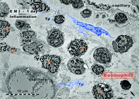
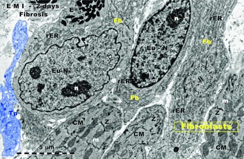
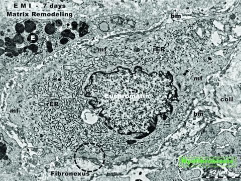
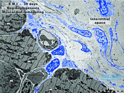
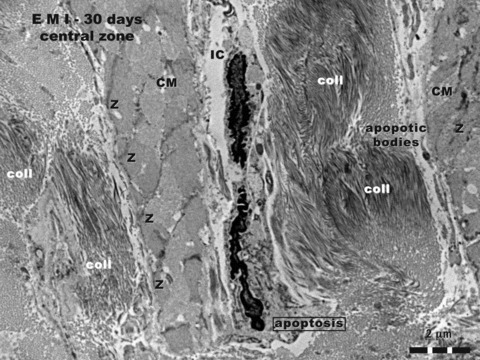
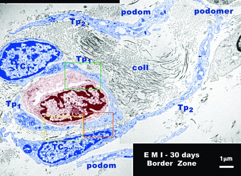
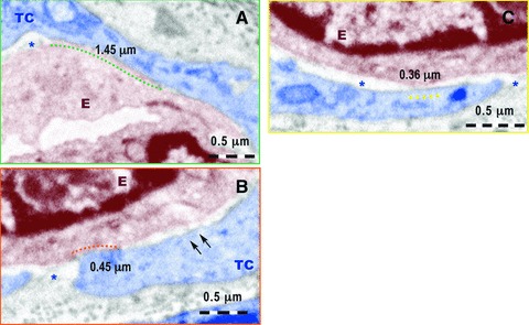
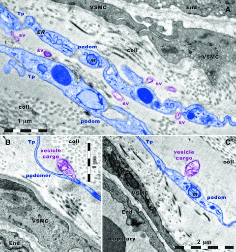
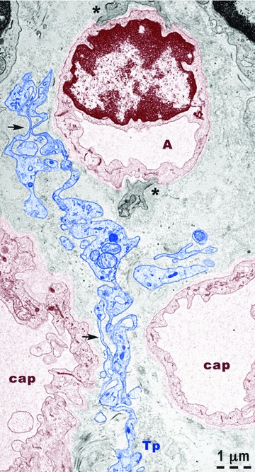
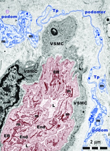
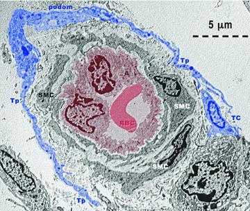
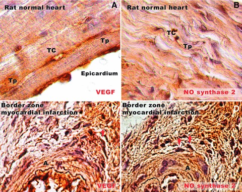
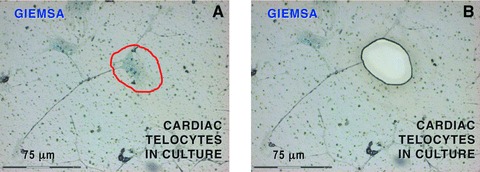
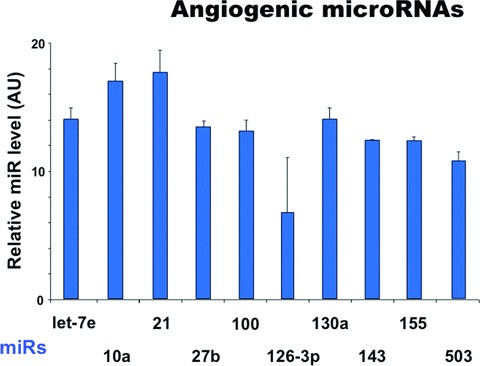
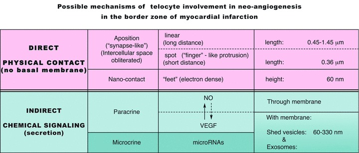
We used rat experimental myocardial infarction to study the ultrastructural recovery, especially neo-angiogenesis in the infarction border zone. We were interested in the possible role(s) of telocytes (TCs), a novel type of interstitial cell very recently discovered in myocardim (see http://www.telocytes.com). Electron microscopy, immunocytochemistry and analysis of several proangiogenic microRNAs provided evidence for TC involvement in neo-angiogenesis after myocardial infarction. Electron microscopy showed the close spatial association of TCs with neoangiogenetic elements. Higher resolution images provided the following information: (a) the intercellular space between the abluminal face of endothelium and its surrounding TCs is frequently less than 50 nm; (b) TCs establish multiple direct nanocontacts with endothelial cells, where the extracellular space seems obliterated; such nanocontacts have a length of 0.4-1.5 μm; (c) the absence of basal membrane on the abluminal face of endothelial cell. Besides the physical contacts (either nanoscopic or microscopic) TCs presumably contribute to neo-angiognesis via paracrine secretion (as shown by immunocytochemistry for VEGF or NOS2). Last but not least, TCs contain measurable quantities of angiogenic microRNAs (e.g. let-7e, 10a, 21, 27b, 100, 126-3p, 130a, 143, 155, 503). Taken together, the direct (physical) contact of TCs with endothelial tubes, as well as the indirect (chemical) positive influence within the 'angiogenic zones', suggests an important participation of TCs in neo-angiogenesis during the late stage of myocardial infarction.


Figures

Fig 1
Fig 1
Rat experimental myocardial infarction. Border zone: 1-day-old. Transmission electron microscopy. The inflammatory granulocyte reaction dominates: granulocyte infiltration, mainly eosinophils (E) and neutrophils (PMN); a mast cell (M) is visible; the arrow indicates an apoptotic cell. A telocyte (TC) and several telopodes (Tp) are digitally blue coloured.
Fig 2
Fig 2
Rat experimental myocardial infarction. Border zone: 2-day-old. Transmission electron microscopy. Three fibroblasts (upper part) intermingle with three cardiomyocytes (lower part). The accumulation of fibroblasts indicates the beginning of fibrosis. Note that usually, in normal myocardium, there are no fibroblast clusters; Fb: fibroblast; rER: rough endoplasmic reticulum; Eu-N: euchromatin; n: nucleolus; CM: cardiomyocyte; m: mitochondria; Z: Z line. Centriole presence (black dotted line) suggests fibroblast mitosis. Tp: telopode (digitally blue coloured); E: eosinophil.
Fig 3
Fig 3
Rat experimental myocardial infarction. Border zone: 7-day-old. Transmission electron microscopy. Note the presence of a typical myofibroblast. Such cells are responsible for matrix remodelling. Myofibroblasts have intermediate features between fibroblasts and smooth muscle cells: prominent rough endoplasmic reticulum (rER, like fibroblasts), and myofilaments (mf, like smooth muscle cells) located under the cell membrane. The characteristic ultrastructural feature, the fibronexus, which is a cell-to-stroma attachment, is highlighted by black dotted circle. E: eosinophil; coll: collagen fibrils.
Fig 4
Fig 4
Rat experimental myocardial infarction. Border zone: 30-day-old. Transmission electron microscopy. This low magnification view shows four cardiomyocytes (CM), two blood capillaries (1 and 2) and numerous telocytes (TC) with long and slender telopodes (Tp). Note the close spatial relationship between TC/Tp and capillary-1 wall (endothelium). Capillary-1 is presumably a neo-capillary created in the interstitial space. Capillary-2, between three cardiomyocytes (CMs), has a TC and Tps in the vicinity, but the distance between abluminal membrane of endothelium and TC/Tp plasma membrane is wider. Thus, capillary-2 is probably an ‘old’ capillary.
Fig 5
Fig 5
Rat experimental myocardial infarction. Central zone of the scar: 30-day-old; transmission electron microscopy. Three structural elements can be recognized in this image: (a) abundant deposits of collagen fibrils (coll), cross or obliquely cut; (b) damaged cardiomyocytes (CM), ‘homogenized’ myofibrils, only positions of Z lines being recognizable; (c) apoptotic interstitial cells (IC), and apoptotic bodies.
Fig 6
Fig 6
Typical echocardiogram of rat experimental myocardial infarction (30-day-old). The black arrow indicates the dyskinetic region of ventricular free wall corresponding to myocardial infarction area. The corresponding ECG in the lower part of the figure shows typical elevation of ST segment, suggestive for acute myocardial infarction.
Fig 7
Fig 7
Rat experimental myocardial infarction. Border zone: 30-day-old. Transmission electron microscopy. A new-formed blood capillary with an anfractuous and narrow lumen is shown (brown colour) in the mass of collagen fibrils (coll) of the scar. This is surrounded by two telocytes (TC1 and TC2 - blue colour) and their corresponding telopodes (TP1 and Tp2). Typically podoms (dilated portions) and the intercalary podomers (thin portions of Tp) can be observed. At the level of podoms there are many mitochondria (m), elements of endoplasmic reticulum and caveolae. Note the close spatial relationships between telopodes and endothelial cells. The space between telopodes and the membrane of endothelial cell is occasionally less than 50 nm and there is no visible endothelial basal lamina.
Fig 8
Fig 8
Higher magnification of the fields marked by colour rectangles in Figure 7. (A) A nanocontact of about 1.45 μm long between the telocyte (TC) and the membrane of the endothelial cell (E) is marked by the green dotted line. The asterisk indicates the intercellular space between TC and E. No endothelial basal lamina is present. (B) A nanocontact of about 0.45 mm long between the E and TC is indicated by the red dotted line. The asterisk mark intercellular space without endothelial basal membrane and the two arrows show possible electron dense ‘feet’ connecting the endothelial cell membrane and the TC membrane. (C) A nanocontact of 0.36 mm long between endothelial cell membrane and TC is indicated by yellow dotted line between the endothelial cell membrane and TC membrane. No basal lamina is visible in the extracellular space between the endothelium and TC (asterisks).
Fig 9
Fig 9
Rat experimental myocardial infarction. Border zone: 30-day-old. Transmission electron microscopy. Telopodes (Tp) are situated in close vicinity of blood vessels. Vascular smooth muscle cells (VSMC) and endothelial cells (End) have many caveolae. Note the alternation of podoms and podomers. Podoms contain: mitochondria (m), and endoplasmic reticulum (ER) and caveolae (cav). (A) Shed vesicles (sv) are released from podoms. A vesicle cargo multivesicular is formed from a podomer (B) and another one is formed from a podom (C). coll: collagen.
Fig 10
Fig 10
Rat experimental myocardial infarction. Border zone: 30-day-old. Transmission electron microscopy. A telopode (Tp - blue colour) is located between three new-formed capillaries (cap). At the level of capillary A, basal lamina and fragments of pericytes (asterisks) are visible. The capillary endothelium (brown colour) has many vesicles of transcytosis. Note the alternation of podoms (containing mitochondria, endoplasmic reticulum and caveolae) and podomers (thin segments) – arrows.
Fig 11
Fig 11
Rat experimental myocardial infarction. Border zone: 30-day-old. Arteriologenesis. Transmission electron microscopy. A telopode (Tp) surrounding a new-formed blood vessel. Tall endothelial cells (End) with numerous mitochondria (m) and endoplasmic reticulum (ER) cisternae, circumvent a narrow and irregular lumen (L). VSMC: vascular smooth muscle cell.
Fig 12
Fig 12
Rat experimental myocardial infarction. Border zone: 30-day-old. Arteriologenesis. Transmission electron microscopy. Telopodes (Tp) with typical podoms and podomers, from at least one telocyte (TC - blue colour) are encircling an arteriole (A: brown colour). Note: (a) the endothelium (brown coloured) surrounding the lumen which contains a red blood cell (RBC: red colour); (b) lamina elastica interna – a clear space surrounding the endothelium; (c) 2–3 layers of smooth muscle cells (SMC).
Fig 13
Fig 13
Rat normal heart tissue (A, B) and border zone of infarcted heart tissue (C, D) in experimental myocardial infarction (30-day-old). (A) Immunostaining for VEGF demonstrates positive expression in the subepicardial area. A silhouette of a telocyte (TC) can be seen parallel with the epicardium. Very long and slender telopodes (Tp) are obvious. (B) Positive expression for NOS2 in rat normal myocardium. A Tp splits dichotomously near the TC cell body, and runs parallel with cardiac fibres. (C) In the border zone of infarcted heart tissue, near a new-formed arteriole (A), a cell (most probable a TC, taking into account the morphology) is positive for VEGF (arrow). (D) Positive reaction for NOS2 in two different cells (arrows) near a new-formed blood vessel in the border zone of infarction. Their prolongations are neighbouring the blood vessel wall. Original magnifications: (A, B) – 100×; (C, D) – 60×.
Fig 14
Fig 14
Laser capture microdissection of cardiac telocytes in culture. Because other interstitial cells than telocytes might be present, only cells with ‘multipolar’ body and at least one prolongation of more than 50 mm long were considered telocytes. Such prolongations had typical telopode conformation, with podoms (dilated portions) and podomers (thin segments). (A) The telocyte body encircled by the red line was dissected out by laser capture microscopy. (B) The corresponding area of Figure 11A after microdissection. A total of 500 telocyte bodies were collected and analysed for microRNAs. 20× objective.
Fig 15
Fig 15
Expression levels of microRNAs up-regulated in telocytes. The relative expression was determined by real-time PCR. RNU43 snoRNA and U6 snRNA were used as normalizing controls. The error bars are mean + S.D. (n = 3) and the data are representative for two replicate experiments. AU: arbitrary units.
Fig 16
Fig 16
Possible mechanisms of telocyte involvement in neo-angiogenesis in the border zone of myocardial infarction.

References

    1. Caulfield J, Klionsky B. Myocardial ischemia and early infarction: an electron microscopic study. Am J Pathol. 1959;35:489–523. - PMC - PubMed
    1. Page E, Polimeni PI. Ultrastructural changes in the ischemic zone bordering experimental infarcts in rat left ventricles. Am J Pathol. 1977;87:81–104. - PMC - PubMed
    1. Gottlieb GJ, Kubo SH, Alonso DR. Ultrastructural characterization of the border zone surrounding early experimental myocardial infarcts in dogs. Am J Pathol. 1981;103:292–303. - PMC - PubMed
    1. Axford-Gatley RA, Wilson GJ. The “border zone” in myocardial infarction. An ultrastructural study in the dog using an electron-dense blood flow marker. Am J Pathol. 1988;131:452–64. - PMC - PubMed
    1. Schaper J. Ultrastructural changes of the myocardium in regional ischaemia and infarction. Eur Heart J. 1986;7B:3–9. - PubMed

Publication types

Substances