Comparative Study
. 2011 Oct;119(5):333-8.
doi: 10.1111/j.1600-0722.2011.00854.x.

Requirement of alveolar bone formation for eruption of rat molars

Affiliations
Comparative Study

Requirement of alveolar bone formation for eruption of rat molars

Gary E Wise et al. Eur J Oral Sci. 2011 Oct.

Abstract

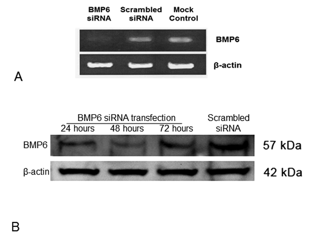
Tooth eruption is a localized event that requires a dental follicle (DF) to regulate the resorption of alveolar bone to form an eruption pathway. During the intra-osseous phase of eruption, the tooth moves through this pathway. The mechanism or motive force that propels the tooth through this pathway is controversial but many studies have shown that alveolar bone growth at the base of the crypt occurs during eruption. To determine if this bone growth (osteogenesis) was causal, experiments were designed in which the expression of an osteogenic gene in the DF, bone morphogenetic protein-6 (Bmp6), was inhibited by injection of the first mandibular molar of the rat with a small interfering RNA (siRNA) targeted against Bmp6. The injection was followed by electroporation to promote uptake of the siRNA. In 45 first molars injected, eruption was either delayed or completely inhibited (seven molars). In the impacted molars, an eruption pathway formed but bone growth at the base of the crypt was greatly reduced compared with the erupted first-molar controls. These studies show that alveolar bone growth at the base of the crypt is required for tooth eruption and that Bmp6 may be essential for promoting this growth.


Conflict of interest statement

Conflicts of interest: Authors declare no conflict of interest.

Figures

Figure 1
Figure 1
In vivo expression of BMP6 in the dental follicles (DF) of the first mandibular molars of postnatal rats as determined by real-time RT-PCR. Chronologically increased expression of BMP6 was seen in the postnatal days examined. Different letters above the bars indicate that the means were significantly different at P≤0.05 in ANOVA and LSD tests. Bars that contain the same letter are not significantly different; e.g., Day 11 (A) and Day 9 (AB) are not significantly different.
Figure 2
Figure 2
Gene knockdown efficiency of siRNA against BMP6 (BMP6-siRNA) was determined by conventional RT-PCR and Western blotting. (A) RT-PCR indicated that BMP6 gene expression in BMP6-siRNA transfected cells was reduced to an almost undetectable level at 48 h after transfection, whereas no notable reduction of BMP6 expression was seen in the cells transfected with scrambled siRNA as compared to the untreated controls. In addition, siRNA transfection appeared to have no effect on expression of the β-actin gene. (B) Western blotting analysis showed that the maximum knockdown effect on BMP6 protein was at 48 h after transfection, but the knockdown effect lasts at least 72 h after transfection.
Figure 3
Figure 3
Transfection of DF with siRNA BMP6 resulted in tooth impaction. (A) Mandible showing the impacted and erupted 1st molars at day 30. Note that the eruption pathway still formed above the impacted molar. (B, C) Histology sections also show the eruption pathway above the impacted molar (B) and that the amount of alveolar bone at the base of the impacted molar was much less (B) as compared to the base of the erupted molar (C).
Figure 4
Figure 4
(A) Comparison of the alveolar bone between the impacted 1st molar and the adjacent erupted 2nd molar at day 30 showing that the impacted 1st molar had less bone at its base than did the base of the erupted 2nd molar. (B &C) High power magnifications of the same area illustrates the differences in the quantity of the alveolar bone (AB) at the base of these molars; i.e., less bone under the 1st molar.
Figure 5
Figure 5
Comparison of the impacted and erupted 1st molars at different postnatal days. A significant reduction of the amount of alveolar bone (AB) was seen at the base of the impacted 1st molars from pups of postnatal days 20 and 30 (A, C) vs. the large amount of bone seen at the base of the erupted 1st molars of the same age (B, D). Note that the alveolar bone (AB) at the base of the erupted molars appeared to be more compact than the bone in the comparable part of the impacted molars (A vs. B; C vs. D).

References

    1. Wise GE, King GJ. Mechanisms of Tooth Eruption and Orthodontic Tooth Movement. J Dent Res. 2008;87:414–434. - PMC - PubMed
    1. Wise GE. Cellular and molecular basis of tooth eruption. Orthod Craniofac Res. 2009;12:67–73. - PMC - PubMed
    1. Cahill DR, Marks SC., Jr Tooth eruption: evidence for the central role of the dental follicle. J Oral Pathol. 1980;9:189–200. - PubMed
    1. Marks SC., Jr Pathogenesis of osteopetrosis in the ia rat: reduced bone resorption due to reduced osteoclast function. Am J Anat. 1973;138:165–189. - PubMed
    1. Cotton WR, Gaines JF. Unerupted dentition secondary to congenital osteopetrosis in the Osborne-Mendel rat. Proc Soc Exp Biol Med. 1974;146:554–561. - PubMed

Publication types

MeSH terms