. 2011 Oct 28;286(43):37483-95.
doi: 10.1074/jbc.M111.284885. Epub 2011 Sep 6.

Modification of histones by sugar β-N-acetylglucosamine (GlcNAc) occurs on multiple residues, including histone H3 serine 10, and is cell cycle-regulated

Affiliations

Modification of histones by sugar β-N-acetylglucosamine (GlcNAc) occurs on multiple residues, including histone H3 serine 10, and is cell cycle-regulated

Suisheng Zhang et al. J Biol Chem. .

Abstract

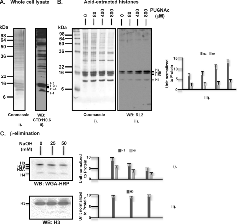
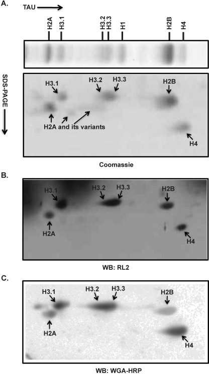
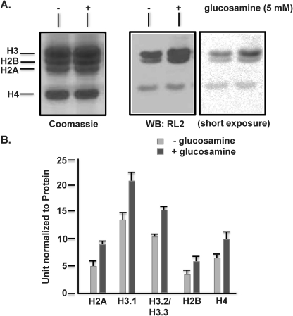
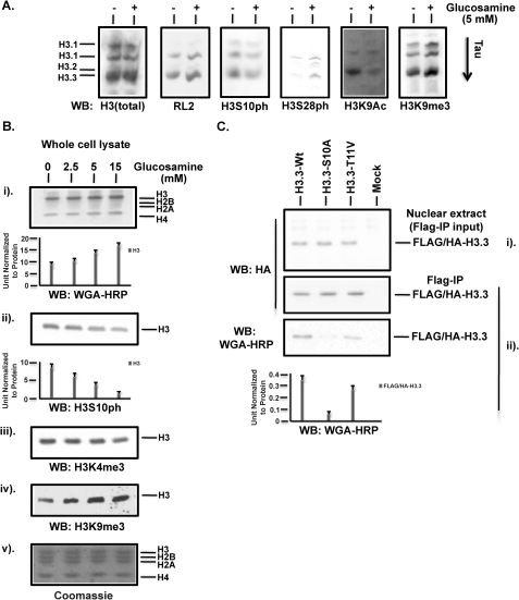
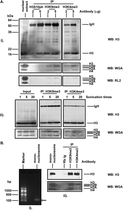
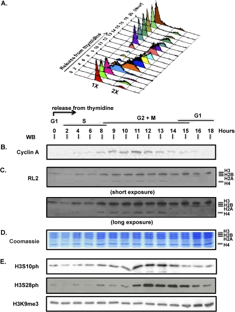
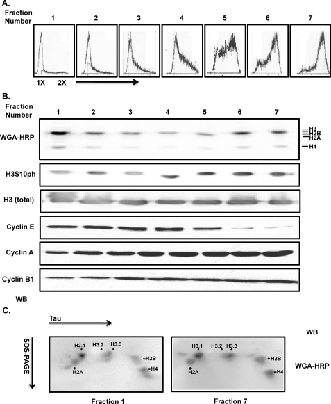
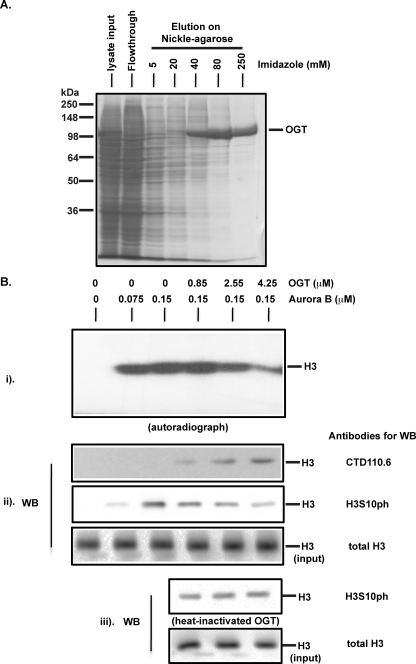
The monosaccharide, β-N-acetylglucosamine (GlcNAc), can be added to the hydroxyl group of either serines or threonines to generate an O-linked β-N-acetylglucosamine (O-GlcNAc) residue (Love, D. C., and Hanover, J. A. (2005) Sci. STKE 2005 312, 1-14; Hart, G. W., Housley, M. P., and Slawson, C. (2007) Nature 446, 1017-1022). This post-translational protein modification, termed O-GlcNAcylation, is reversible, analogous to phosphorylation, and has been implicated in many cellular processes. Here, we present evidence that in human cells all four core histones of the nucleosome are substrates for this glycosylation in the relative abundance H3, H4/H2B, and H2A. Increasing the intracellular level of UDP-GlcNAc, the nucleotide sugar donor substrate for O-GlcNAcylation enhanced histone O-GlcNAcylation and partially suppressed phosphorylation of histone H3 at serine 10 (H3S10ph). Expression of recombinant H3.3 harboring an S10A mutation abrogated histone H3 O-GlcNAcylation relative to its wild-type version, consistent with H3S10 being a site of histone O-GlcNAcylation (H3S10glc). Moreover, O-GlcNAcylated histones were lost from H3S10ph immunoprecipitates, whereas immunoprecipitation of either H3K4me3 or H3K9me3 (active or inactive histone marks, respectively) resulted in co-immunoprecipitation of O-GlcNAcylated histones. We also examined histone O-GlcNAcylation during cell cycle progression. Histone O-GlcNAcylation is high in G(1) cells, declines throughout the S phase, increases again during late S/early G(2), and persists through late G(2) and mitosis. Thus, O-GlcNAcylation is a novel histone post-translational modification regulating chromatin conformation during transcription and cell cycle progression.


Figures

FIGURE 1.
FIGURE 1.
Human histones are post-translationally modified by glycosylation, specifically O-GlcNAcylation. A, whole cell lysates were prepared from cultured HEK293 cells that were immediately denatured in SDS-PAGE sampling buffer at 95 °C for 5 min. After electrophoresis through a 15% SDS-polyacrylamide gel, Western blotting (WB) was performed with the mouse monoclonal antibody CTD110.6, specific to O-GlcNAc. B, histones were prepared by acid extraction from HEK293 cells as described under “Experimental Procedures.” O-(2-Acetamido-2-deoxy-d-glucopyranosylidene) amino N-phenylcarbamate (PUGNAc) was included at the indicated concentrations in all the buffers to inhibit O-GlcNAcase that removes O-GlcNAc. After SDS-PAGE of extracted histones, Western blotting was performed with the mouse monoclonal antibody RL2, also specific to O-GlcNAc, which is quantified three times via densitometry and presented in units normalized to the protein levels, the errors indicating means ± S.D. C, histone O-GlcNAcylation was examined for its sensitivity to mild base treatment leading to β-elimination of the O-linked GlcNAc. For this purpose, the acid-extracted histones were incubated without or with 25 and 50 mm NaOH for 30 min at 37 °C, followed by SDS-PAGE and Western blotting with a lectin probe WGA-HRP that is also specific to O-GlcNAc. A rabbit polyclonal antibody against total histone H3 was used for presenting an input control. Quantifications are presented as above.
FIGURE 2.
FIGURE 2.
Histone O-GlcNAcylation is further resolved using two-dimensional gel electrophoresis. Acid-extracted histones from HEK293 cells were separated first by TAU gel electrophoresis, followed by a second dimensional separation through 15% SDS-PAGE. Histones after TAU or two-dimensional gel electrophoresis were visualized by Coomassie Blue (A) or transferred to a nitrocellulose membrane for Western blot (WB) with the antibody RL2 (B) or WGA-HRP (C), both of which revealed the levels of O-GlcNAcylation of individual histones and some of their isoforms or variants, as indicated.
FIGURE 3.
FIGURE 3.
An in vivo treatment with glucosamine-enhanced global levels of histone O-GlcNAcylation. A, HEK293 cells in culture were treated by glucosamine at 5 mm for 16 h, followed by acid-extraction of histones, SDS-PAGE, and Western blot (WB) with antibody RL2 to examine histone O-GlcNAcylation. B, acid-extracted histones as shown in A were separated in parallel by two-dimensional gel electrophoresis as shown in Fig. 2, followed by Western blot with WGA-HRP for detection of individual histone O-GlcNAcylation. Quantifications based on densitometry were presented for a comparison of individual histone O-GlcNAcylation without or with the glucosamine treatment. Quantifications, in units normalized to the protein levels, are the average and the standard deviation of three experiments.
FIGURE 4.
FIGURE 4.
Histone H3 serine 10 is a site of O-GlcNAcylation. A, enhancement of histone O-GlcNAcylation by glucosamine is associated with a decreased level of phosphorylation of H3S10 (H3S10ph). Acid-extracted histones from HEK293 cells untreated (−) or treated (+) with glucosamine were separated by TAU gel electrophoresis, followed by Western blotting (WB) with antibody RL2 to detect histone O-GlcNAcylation and antibodies against various histone H3 post-translational modifications, as indicated, as well as an antibody against total H3. B, Western blot of whole cell lysates after glucosamine treatment at the indicated concentrations. Quantifications based on densitometry are presented in units normalized to the protein levels and are the average and the standard deviations of three experiments. SDS-PAGE followed by Coomassie staining indicates total histones. C, FLAG- and HA-tagged wild-type H3.3, its S10A and T11V mutant expressed from plasmid pOZ-FH-C transfected into HEK293 cells. Cells at 48 h after transfection were collected for preparing the nuclear extracts (panel i) and then immunoprecipitates (panel ii) with a mouse monoclonal antibody (M2) against the FLAG tag. Recombinant H3.3 proteins were examined by Western blot with WGA-HRP for detecting O-GlcNAc from recombinant H3.3. The WGA signals were quantified and normalized to the signals of anti-HA tag antibody examining recombinant H3.3 in the FLAG tag pulldown.
FIGURE 5.
FIGURE 5.
O-GlcNAcylation is associated with both inactive and active marks of histone H3. A, panel i, histone immunoprecipitations (IP) using HEK293 nuclear extracts were performed with increasing amounts of antibody against H3S10p, H3K9me3, and H3K4me3 as indicated. Immunoprecipitated histone H3 was examined by Western blot (WB) with the antibody against total H3, WGA-HRP, or antibody RL2 to detect histone O-GlcNAcylation in the immunoprecipitates. Panel ii, histone immunoprecipitations as above and as indicated were performed with nuclear extracts sonicated for indicated number of times, followed by Western blot of immunoprecipitated histone H3 and histone O-GlcNAcylation revealed by WGA-HRP. B, immunoprecipitation of mononucleosomes derived from micrococcal nuclease digestion of the HEK293 nuclei (panel i) and Western blot showing histone O-GlcNAcylation associated with mononucleosomes immunoprecipitated with the indicated antibodies (panel ii). Rb lg is rabbit immunoglobulin.
FIGURE 6.
FIGURE 6.
Histone O-GlcNAcylation is cell cycle-regulated as assessed by double thymidine block and release. HeLa cells were synchronized by a double treatment with thymidine to block cell cycle at the transition of G1 to S phase. Cell cycle was followed at the indicated time points after release from the thymidine block. A, FACS profiles showing a progression through the cell cycle. B, Western blot (WB) using whole cell lysates and an antibody against cyclin A. Cell cycle phases estimated from the FACS data, cyclin A, and H3S10ph (see below) are indicated above the cyclin A blot. C, histone O-GlcNAcylation examined with the antibody RL2. D, histones visualized by Coomassie-staining and SDS-PAGE. E, Western blot showing the histone H3 post-translational modifications H3S10ph, H3S28ph, and H3K9me3. Coomassie-stained total histones and H3K9me3 serve as loading controls.
FIGURE 7.
FIGURE 7.
Histone O-GlcNAcylation is cell cycle-regulated as assessed by centrifugal elutriation. Asynchronously growing human acute T lymphoblastic leukemia cells, CCRF-CEM, were loaded into an elutriator rotor, and the flow rate was increased in steps to elute small to large cells. A, FACS analysis of the seven eluted fractions indicates successful cell cycle fractionation. B, Western blot (WB) analyses of the indicated proteins after SDS-PAGE. Histones were prepared by acid extraction and detected using WGA-HRP and antibodies against H3S10ph, total H3, cyclin E, cyclin A, and cyclin B1 as indicated. C, O-GlcNAcylation of histones prepared from G1 (fraction 1) and G2 + M (fraction 7) cells assessed by two-dimensional gel electrophoresis and Western blotting with WBA-HRP. Note that the faint spot below and to the left of the H2A spot that is visible in fraction 1 is normally not visible in these two-dimensional gels (24) and is either a gel artifact or possibly a minor histone variant specific to G1.
FIGURE 8.
FIGURE 8.
OGT suppressed phosphorylation of histone H3 at serine 10 by Aurora B in vitro. A, purification of baculovirus-expressed OGT on Ni2+-agarose. Proteins from whole lysate, flow-through, and fractions eluted at the indicated concentrations of imidazole were examined by SDS-PAGE followed by Coomassie stain. B, effect of OGT on phosphorylation of histone H3 by Aurora B. For all the reactions, UDP-GlcNAc was included in the assay mixtures as described under “Experimental Procedures” (panel i). Aurora B kinase assay performed with [γ-32P]ATP as the phosphate donor. Panel ii, Aurora B kinase assay performed with unlabeled ATP (cold) and phosphorylation examined by Western blotting (WB) with antibody specifically recognizing phosphorylated serine 10 of H3. The O-GlcNAcylation of histone H3 by OGT was examined with the anti-O-GlcNAc antibody CTD110.6. Histone H3 as substrate in the assays was presented by Western blot with an antibody against unmodified H3. Panel iii, as a control of OGT activity, the protein was heated at 95 °C for 5 min before addition to the assay mixtures.

Similar articles

Cited by

References

    1. Love D. C., Hanover J. A. (2005) Sci. STKE 2005, re13 - PubMed
    1. Hart G. W., Housley M. P., Slawson C. (2007) Nature 446, 1017–1022 - PubMed
    1. Zachara N. E., Hart G. W. (2004) Trends Cell Biol. 14, 218–221 - PubMed
    1. Hurtado-Guerrero R., Dorfmueller H. C., van Aalten D. M. (2008) Curr. Opin. Struct. Biol. 18, 551–557 - PubMed
    1. Shafi R., Iyer S. P., Ellies L. G., O'Donnell N., Marek K. W., Chui D., Hart G. W., Marth J. D. (2000) Proc. Natl. Acad. Sci. U.S.A. 97, 5735–5759 - PMC - PubMed

Publication types

MeSH terms

LinkOut - more resources