. 2011 Oct 28;286(43):37094-107.
doi: 10.1074/jbc.M111.242438. Epub 2011 Sep 6.

A unique role for heat shock protein 70 and its binding partner tissue transglutaminase in cancer cell migration

Affiliations

A unique role for heat shock protein 70 and its binding partner tissue transglutaminase in cancer cell migration

Lindsey K Boroughs et al. J Biol Chem. .

Abstract

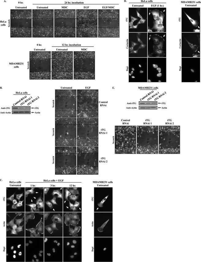
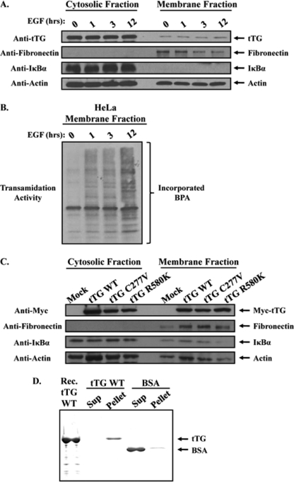
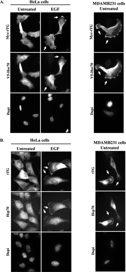
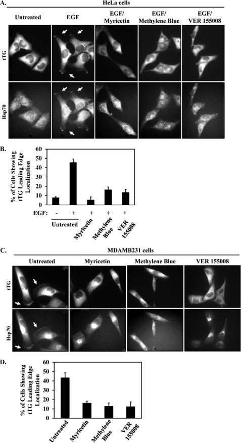
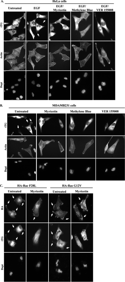
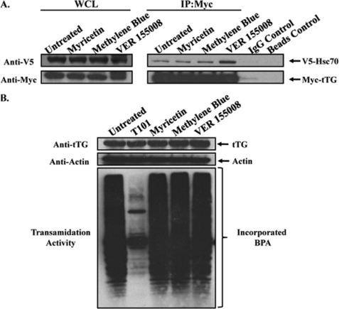
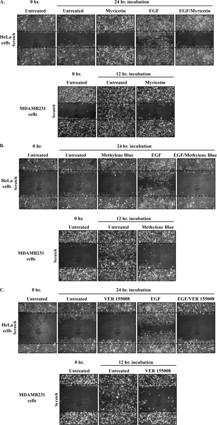
Cell migration is essential for several important biological outcomes and is involved in various developmental disorders and disease states including cancer cell invasiveness and metastasis. A fundamental step in cell migration is the development of a leading edge. By using HeLa carcinoma cells as an initial model system, we uncovered a surprising role for the heat shock protein 70 (Hsp70) and its ability to bind the protein cross-linking enzyme, tissue transglutaminase (tTG), in cancer cell migration. Treatment of HeLa cells with EGF results in the activation of a plasma membrane-associated pool of tTG and its redistribution to the leading edges of these cells, which are essential events for EGF-stimulated HeLa cell migration. However, we then found that the ability of tTG to be localized to the leading edge is dependent on Hsp70. Similarly, the localization of tTG to the leading edges of MDAMB231 breast carcinoma cells, where it also plays an essential role in their migration, has a strict requirement for Hsp70. Treatment of these different cell lines with inhibitors against the ATP hydrolytic activity of Hsp70 prevented tTG from localizing to their leading edges and thereby blocked EGF-stimulated HeLa cell migration, as well as the constitutive migration normally exhibited by MDAMB231 cells. These findings highlight a new and unconventional role for the chaperonin activity of Hsp70 in the localization of a key regulatory protein (tTG) at the leading edges of cancer cells and the important consequences that this holds for their ability to migrate.


Figures

FIGURE 1.
FIGURE 1.
tTG is localized to the leading edges of actively migrating cells, and its cross-linking activity is necessary for cell migration. A, scratch assays were performed on HeLa cells (top panels) and MDAMB231 cells (bottom panels) treated without (Untreated) or with EGF and without or with MDC as indicated. MDAMB231 cells were fixed 12 h after striking the wound; HeLa cells were fixed after 24 h. The cells were then visualized using light microscopy, and the extent of wound closure was determined. One set of untreated cells was fixed immediately after striking the wound (Untreated 0 h.) to indicate the size of the initial wounds. The widths of the initial wounds are indicated by dashed lines. B, the extracts collected from HeLa cells transfected with control-RNAi, tTG-RNAi 1, or tTG-RNAi 2 were immunoblotted with tTG and actin antibodies (left panels). Scratch assays were then performed on cells transfected with the same siRNAs, treated without (Untreated) or with EGF. The cells were processed as outlined in A (right panels). C and D, duplicate sets of serum-starved cultures of HeLa cells and MDAMB231 cells were treated without (Untreated) or with EGF for increasing lengths of time, as indicated, and then were fixed. C, immunofluorescence was performed on one set of the cells using a tTG antibody, rhodamine-conjugated phalloidin (Actin), and DAPI (to stain nuclei). D, immunofluorescence was performed on the second set of cells using tTG and cortactin antibodies and DAPI. Representative fluorescent images of the cells are shown, and the localization of tTG and cortactin at leading edges is indicated with arrows. E, the extracts collected from MDAMB231 cells transfected with control-RNAi, tTG-RNAi 1, or tTG-RNAi 2 were immunoblotted with tTG and actin antibodies (top panels). Scratch assays were then performed on cells transfected with the same siRNAs, and the cells were processed as outlined in A (bottom panels).
FIGURE 2.
FIGURE 2.
A pool of tTG is constitutively associated with the plasma membrane. Serum-starved HeLa cells that had been treated without (lane 0) or with EGF for increasing lengths of time, as indicated, were homogenized and then subjected to differential centrifugation to separate cytosolic and membrane fractions. A, the cellular fractions were immunoblotted with tTG, fibronectin, IκBα, and actin antibodies. B, the same membrane fractions were also assayed for tTG transamidation activity by determining the incorporation of BPA into lysate proteins. C, actively growing HeLa cells that had been either mock transfected without DNA (Mock) or transfected with various Myc-tagged forms of tTG including wild-type (tTG WT), a transamidation-defective mutant (tTG C277V), or a GTP-binding-defective mutant (tTG R580K) were homogenized and then subjected to differential centrifugation to isolate cytosolic and membrane fractions. The fractions were immunoblotted with Myc, fibronectin, IκBα, and actin antibodies. D, synthetic liposomes were prepared by extrusion, and then equal amounts of this preparation were combined with either 5 μg of recombinant tTG (tTG WT) or 5 μg of BSA. After a 15-min incubation, the liposomes were pelleted by centrifugation, and the resulting supernatant (Sup) and liposome (Pellet) fractions were resolved by SDS-PAGE. The gel was then stained with Quick Blue to detect the proteins. A lane containing recombinant tTG (Rec. tTG WT) was included as a standard.
FIGURE 3.
FIGURE 3.
Hsp70 interacts with plasma membrane-associated tTG. A, serum-starved HeLa cells that had been treated without (lane 0) or with EGF for 12 h were homogenized and then subjected to differential centrifugation to isolate the membrane components of the cells. Immunoprecipitations with a tTG antibody (IP:tTG) were performed on the membrane extracts, and the resulting immunocomplexes were resolved by SDS-PAGE. The gel was then stained with Colloidal Blue to detect the proteins that co-immunoprecipitated with tTG. One protein band (Mr = ∼70 kDa), denoted with an arrow, was determined to contain two isoforms of the heat shock protein 70 family, Hsp70 and Hsc70, by mass spectrometry. B, the protein sequences of human Hsp70 and Hsc70 are shown in gray. The peptide fragments identified by mass spectrometry are shaded in black. C, immunoprecipitations with a Myc antibody were performed on the extracts of HeLa cells ectopically expressing V5-tagged Hsc70 and Myc-tagged tTG, treated without (Untreated) or with EGF. The whole cells lysates (WCL) and the resulting immunocomplexes (IP:Myc) were immunoblotted with V5 and Myc antibodies. Nonspecific mouse IgG control antibody and beads-only control immunoprecipitations were also performed on the extracts to show that the Hsc70-tTG interaction was specific. D, immunoprecipitations with a Myc antibody were performed on the extracts of actively growing HeLa (top panels) and MDAMB231 cells (bottom panels) that were ectopically expressing V5-tagged Hsc70 and a Myc-tagged form of either wild-type tTG (tTG WT) or a transamidation-defective form of tTG (tTG C277V). The whole cell lysates (WCL) and the resulting immunocomplexes (IP:Myc) were immunoblotted with V5 and Myc antibodies. Nonspecific mouse IgG control antibody and beads-only control immunoprecipitations were performed on the extracts collected from HeLa and MDAMB231 cells expressing V5-Hsc70 and Myc-tTG WT to show that the Hsc70-tTG interaction was specific.
FIGURE 4.
FIGURE 4.
tTG and Hsp70 co-localize to the leading edges of cells. A, HeLa and MDAMB231 cells ectopically expressing V5-Hsc70 and Myc-tTG were fixed and then subjected to immunofluorescence using V5 and Myc antibodies and DAPI (to stain nuclei). Representative images of the transfectants are shown, with the co-localization of the ectopically expressed forms of tTG and Hsc70 at leading edges being highlighted with arrows. B, serum-starved HeLa cells and MDAMB231 cells were treated without (Untreated) or with EGF for 12 h, as indicated, and fixed. Immunofluorescence was performed on the cells using tTG and Hsp70 antibodies and DAPI. Representative images of the cells are shown, with the co-localization of tTG and Hsp70 at leading edges being indicated with arrows.
FIGURE 5.
FIGURE 5.
Inhibiting Hsp70 activity blocks the ability of tTG and Hsp70 to localize at leading edges. A, serum-deprived HeLa cells were treated without (Untreated) or with EGF, with or without myricetin, methylene blue, or VER 155008, and then immunofluorescence was performed on the cells using tTG and Hsp70 antibodies. The resulting fluorescent images are shown with the tTG and Hsp70 at leading edges being indicated with arrows. B, quantification of the cells shown in A with tTG at their leading edges. C, serum-starved MDAMB231 cells were treated without (Untreated) or with myricetin, methylene blue, or VER 155008, and then immunofluorescence was performed on the cells using tTG and Hsp70 antibodies. The resulting fluorescent images are shown with the tTG and Hsp70 at leading edges being indicated with arrows. D, quantification of the cells shown in C with tTG at their leading edges. Three independent experiments were performed, with at least 250 cells being scored for each condition. The results from the experiments were then averaged together and graphed. The error bars indicate standard deviation.
FIGURE 6.
FIGURE 6.
Inhibition of Hsp70 does not have a global effect on leading edge proteins. A, serum-deprived HeLa cells were treated without (Untreated) or with EGF, with or without myricetin, methylene blue, or VER 155008, and then immunofluorescence was performed on the cells using a tTG antibody, rhodamine-conjugated phalloidin (Actin), and DAPI. The resulting fluorescent images are shown with the tTG at leading edges being indicated with arrows. B, serum-starved MDAMB231 cells were treated without (Untreated) or with myricetin, methylene blue, or VER 155008, and then immunofluorescence was performed on the cells using a tTG antibody, rhodamine-conjugated phalloidin (Actin), and DAPI. The resulting fluorescent images are shown with the tTG at leading edges being indicated with arrows. C, MDAMB231 cells were transiently transfected with either of two HA-tagged constructs: constitutively active Rac (F28L) or GTP hydrolysis-defective Ras (G12V), and immunofluorescence was performed using HA and tTG antibodies and DAPI. The resulting fluorescent images are shown with the Rac, Ras, and tTG at leading edges being indicated with arrows.
FIGURE 7.
FIGURE 7.
Inhibiting the ATP hydrolytic activity of Hsp70 has no effect on the interaction between tTG and Hsp70, or on the protein cross-linking activity of tTG. A, HeLa cells ectopically expressing V5-Hsc70 and Myc-tTG were treated without (Untreated) or with myricetin, methylene blue, or VER 155008, as indicated, and lysed. Immunoprecipitations (IP) with a Myc antibody were performed on the cell extracts, followed by SDS-PAGE and Western blot analysis using V5 and Myc antibodies. Nonspecific mouse IgG control antibody and beads-only control immunoprecipitations were performed on the extracts from the untreated cells to show that the Hsc70-tTG interaction was specific. B, serum-starved cultures of MDAMB231 cells were treated without (Untreated) or with T101, myricetin, methylene blue, or VER 155008 and then lysed. The extracts were immunoblotted with tTG and actin antibodies (top panels) and assayed for transamidation activity as read-out by the incorporation of BPA into lysate proteins (bottom panel).
FIGURE 8.
FIGURE 8.
Inhibition of Hsp70 blocks the migration of HeLa and MDAMB231 cells. A, scratch assays were performed on serum-deprived HeLa cells treated without (Untreated) or with EGF, with or without myricetin, and on serum-starved MDAMB231 cells with or without myricetin. The MDAMB231 cells were fixed 12 h after striking the wound; HeLa cells were fixed after 24 h. The cells were then visualized using light microscopy, and the extent of wound closure was determined. One set of untreated cells was fixed immediately after striking the wound (Untreated 0 h.) to indicate the size of the initial wounds. The widths of the initial wounds are indicated by dashed lines. B, scratch assays were performed on the cell lines using methylene blue as outlined in A. C, scratch assays were performed on the cell lines using VER 155008 as outlined in A.

References

    1. Raja, Sivamani K., Garcia M. S., Isseroff R. R. (2007) Front. Biosci. 12, 2849–2868 - PubMed
    1. Cyster J. G. (2005) Annu. Rev. Immunol. 23, 127–159 - PubMed
    1. Aman A., Piotrowski T. (2010) Dev. Biol. 341, 20–33 - PubMed
    1. Luster A. D., Alon R., von Andrian U. H. (2005) Nat. Immunol. 6, 1182–1190 - PubMed
    1. Keller R. (2005) Curr. Opin. Cell Biol. 17, 533–541 - PubMed

Publication types

LinkOut - more resources