. 2011 Nov 4;286(44):38768-38782.
doi: 10.1074/jbc.M111.234161. Epub 2011 Sep 5.

Genomic analysis reveals a novel nuclear factor-κB (NF-κB)-binding site in Alu-repetitive elements

Affiliations

Genomic analysis reveals a novel nuclear factor-κB (NF-κB)-binding site in Alu-repetitive elements

Athina Antonaki et al. J Biol Chem. .

Abstract

The transcription factor NF-κB is a critical regulator of immune responses. To determine how NF-κB builds transcriptional control networks, we need to obtain a topographic map of the factor bound to the genome and correlate it with global gene expression. We used a ChIP cloning technique and identified novel NF-κB target genes in response to virus infection. We discovered that most of the NF-κB-bound genomic sites deviate from the consensus and are located away from conventional promoter regions. Remarkably, we identified a novel abundant NF-κB-binding site residing in specialized Alu-repetitive elements having the potential for long range transcription regulation, thus suggesting that in addition to its known role, NF-κB has a primate-specific function and a role in human evolution. By combining these data with global gene expression profiling of virus-infected cells, we found that most of the sites bound by NF-κB in the human genome do not correlate with changes in gene expression of the nearby genes and they do not appear to function in the context of synthetic promoters. These results demonstrate that repetitive elements interspersed in the human genome function as common target sites for transcription factors and may play an important role in expanding the repertoire of binding sites to engage new genes into regulatory networks.


Figures
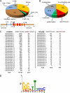
FIGURE 1.
FIGURE 1.
Statistical analysis of p65-bound genomic loci. A, the 366 clones were classified by locations relative to the nearest neighbor gene. The pie chart shows the distribution of p65-binding sites located in promoters (−10 kb to the transcription start site), exons, introns, 5′- and 3′-untranslated regions, remote sites (−10 to −100 kb), and intergenic sites (>100 kb away from genes). Putative NF-κB-binding sites were detected using the TESS and TRANSFAC 7.0 platform. B, pie chart illustrating the number of mismatches of the identified κB sites relative to the consensus κB site. The GGGTTTCACC element, although it bears two mismatches, was classified separately. C, shown is the frequency of the GGGTTTCACC element in human and mouse chromosomes. The distribution of the mutant control sequence GGGTATCACC is also shown for both mouse and human chromosomes. D, position weight matrix of the κB sites derived from the ChIP cloning experiments using THEME.
FIGURE 2.
FIGURE 2.
Genome-wide interaction of NF-κB to selected Alu repeats. A, alignment of 30-bp-long insertions of Alu repeat element family sequences flanking the Alu-κB site. Sequences were retrieved from the NCBI data base and aligned by the Clustal algorithm. The Alu-κB element (5′-GGGTTTCACC-3′) is boxed. B, chromatin prepared from HeLa cells that were either mock-infected (0 h) or infected for 6 h with Sendai virus was immunoprecipitated with the anti-p65 antibody. The presence of each Alu repeat family in the immunoprecipitated material was detected by PCR. C, pie chart illustrating the distribution of the Alu-κB elements found in our clones located in promoters (−10 kb to the transcription start site), exons, introns, 5′- and 3′-untranslated regions, remote sites (−10 to −100 kb), and intergenic sites (>100 kb away from genes).
FIGURE 3.
FIGURE 3.
Recruitment of NF-κB at various non-promoter locations in the genome validates the ChIP-cloning analysis. A, individual ChIP assays of NF-κB binding to randomly selected clones derived from the ChIP-cloning method using chromatin prepared from HeLa cells that were either mock- or virus-infected for 6 h (lanes 1–4) or TNF-α-treated for 1 h (lanes 7 and 8). The chromatin was immunoprecipitated with either the p65 (lanes 1, 2, 7, and 8) or the HMGI(Y) antibody (lanes 3 and 4). The IFN-β promoter was used as a positive control for virus infection and the IL-8 promoter for TNF-α induction. The relative location of the selected clones and the identity of their κB element are indicated at the left. B, same as in A except that the chromatin used was prepared from IB4 lymphoblastoid cells, and the antibodies used were against p52, RelB, and p65. The ELC promoter was used as a positive control for the anti-p52 and anti-RelB ChIPs, and the IL-8 promoter was used as a positive control for the anti-p65 ChIP. Clones have been selected randomly from the anti-p52-ChIP library
FIGURE 4.
FIGURE 4.
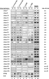
NF-κB induces the recruitment of histone acetylases and general transcription factors to non-promoter regions. Chromatin was prepared from HeLa cells that were either mock-infected (0 h) or infected for 6 h with Sendai virus. The chromatin was immunoprecipitated with the anti-AcH4, anti-AcH3, anti-PolII, and anti-TBP antibodies, and the precipitated DNA fragments were identified by PCR using specific sets of primers corresponding to randomly selected clones.
FIGURE 5.
FIGURE 5.
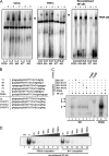
Interaction of NF-κB with the Alu-κB element in vitro. A, an EMSA using radiolabeled oligonucleotide probes derived from randomly selected clones and nuclear extracts from untreated (−TNF) or TNFα-treated (+TNF) HeLa cells (lanes 1–10) or recombinant NF-κB(p50/p65) (lanes 11–20). B, sequence of the probes and the competitor oligonucleotides used in the EMSA experiments. C, EMSA supershift experiments, using the Alu-κB clone 31 as a probe and nuclear extracts prepared from TNF-α-treated HeLa cells. The specificity of the NF-κB-containing complexes was verified by competition (lanes 3–6) and by incubation of the complexes with the anti-p65 (lane 7) and anti-p50 antibody (lane 8). The NF-κB-containing complexes are labeled as a, and the supershifted complexes are labeled as c and b. D, shown is an EMSA experiment using the Alu-κB element derived from clone 11 as a probe and recombinant NF-κB (p50/p65 heterodimer). The DNA-binding reactions were carried out in the absence (lane 1) or in the presence of cold PRDII (lanes 3–8) or Alu-κB clone 11 competitors at the indicated molar ratios.
FIGURE 6.
FIGURE 6.
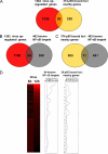
Comparison of genome-wide p65 chromatin occupancy with virus-induced gene expression. A, comparison of the 1,262 genes induced by virus infection with the 374 genes located near the 366 p65-bound loci as displayed in Venn diagrams. The number of genes identified in each case is indicated. B, Venn diagram comparing the number of virus-inducible genes identified in our study with all previously known NF-κB target genes obtained from the Boston University NF-κB Transcription Factors Web site. C, Venn diagram comparing all known NF-κB target genes (462 genes) with the 374 genes located near the 366 p65-bound loci identified by our ChIP-cloning approach. D, heat map showing the expression profile of virus-induced genes at 6 and 12 h postinfection. Red or black represents increase or no change in gene expression relative to uninfected cells, respectively. Only genes displaying significant changes in expression levels (>1.5-fold or 1/1.5-fold; false discovery rate <5%) as compared with the uninfected control in three biological replicates are shown. The moving average of genes in a window of 60 genes that are known to be regulated by NF-κB is shown on the left. The right part illustrates a similar analysis performed with the 64 and 36 genes identified by our study to contain an NF-κB site and to be activated by virus infection. The straight line represents the expected percentage of NF-κB-regulated genes. The group of 64 genes corresponds to previously known NF-κB targets that were identified in our DNA microarray analysis to be induced by virus infection, whereas the group of 36 genes corresponds to genes identified by our ChIP cloning analysis and shown to be induced by virus infection.
FIGURE 7.
FIGURE 7.
Transcriptional properties of genomic p65-bound loci. A, HeLa cells were cotransfected with reporter constructs bearing randomly selected p65-bound loci upstream of the thymidine kinase (TK) minimal promoter directing the expression of luciferase with or without a p65 expression vector. The cells were either mock- or virus-infected for 12 h, and the luciferase activity was determined and plotted after normalization with lacZ. B, Western blotting using some of the extracts generated in Fig. 7A reacted with the anti-p65 antibody (sc-109, Santa Cruz Biotechnology, Inc.). An anti-β-actin antibody (sc-1616, Santa Cruz Biotechnology, Inc.) was used to detect β-actin as an internal control. C, HeLa cells were cotransfected with reporter constructs bearing one, two, or four copies of the GGGTTTCACC or PRDII (GGGAAATTCC) element cloned immediately upstream of the IFN-β minimal promoter in the presence or the absence of the p65 expression vector. The cells were either mock- or virus-infected for 12 h before the luciferase activity was determined. D, shown is an in vitro transcription assay using HeLa nuclear extracts and templates bearing four copies of the PRDII element (PRDIIx4LUC) or four copies of the Alu-κB element (−40LUC #11×4 and −40LUC #39×4). The sequence of the κB oligonucleotides used to generate the templates is shown in Fig. 5B. The in vitro transcription reactions were carried out in the absence or the presence of saturated amounts of recombinant NF-κB according to the scheme shown at the left of the gel. The NF-κB-dependent transcripts generated in the absence or presence of Sarkosyl (an inhibitor of the reinitiation of transcription) are indicated by an arrow (top) and were quantified by the ImageQuant software (Molecular Dynamics) to calculate the rounds of transcription that took place (bottom). A representative experiment of two is shown. Error bars, S.D.
FIGURE 8.
FIGURE 8.
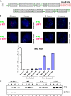
Distinct functional properties of Alu-κB repeat elements. A, maximum DNA sequence alignment between the chip-cloning clone 11 and the 4C-ChIP clone 21. Identical sequences are boxed, and the Alu-κB element is indicated. B, DNA FISH analysis examining interchromosomal interactions between IFN-β and Alu-κB clones. A digoxygenin-labeled bacterial artificial chromosome DNA probe was used to detect the endogenous IFN-β locus and was visualized with FITC-conjugated anti-DIG antibody (green). Biotin-labeled bacterial artificial chromosome DNA probes were used to detect the endogenous Alu-κB clone 21 and 11 loci, as indicated in the figure (top), and visualized with Alexa 568-conjugated streptavidin (red). The stably integrated plasmids bearing clones 11 and 21 were detected through the preparation of plasmid DNA (pKS) biotin-labeled probes (bottom). The circle indicates the colocalized alleles observed only in virus-infected cells. The bottom of B shows a diagrammatic representation of the percentage (mean ± S.D. (error bars)) of cells with colocalization between IFN-β and each of the Alu-κB clones in mock- and virus-infected cells. C, HeLa cells stably transfected with Bluescript-based constructs bearing the 4C-ChIP clone 21 and the ChIP-cloning clone 11 along with untransfected HeLa cells or HeLa cells bearing the empty vector were either mock-infected or infected with Sendai virus for 4, 6, or 8 h, and the isolated RNAs were used as templates for RT-PCR analysis using primers specific for IFN-β. The PCR products were detected by gel electrophoresis.

References

    1. Vallabhapurapu S., Karin M. (2009) Annu. Rev. Immunol. 27, 693–733 - PubMed
    1. Hayden M. S., Ghosh S. (2008) Cell 132, 344–362 - PubMed
    1. Li Q., Verma I. M. (2002) Nat. Rev. Immunol. 2, 725–734 - PubMed
    1. Wan F., Lenardo M. J. (2009) Cold Spring Harb. Perspect. Biol. 1, a000067 - PMC - PubMed
    1. Bonizzi G., Karin M. (2004) Trends Immunol. 25, 280–288 - PubMed

Publication types

LinkOut - more resources