Review
. 2011 Sep;140(3):768-774.
doi: 10.1378/chest.10-2132.

Alternatively activated macrophages and airway disease

Affiliations
Review

Alternatively activated macrophages and airway disease

Derek E Byers et al. Chest. 2011 Sep.

Abstract

Macrophages are the most abundant immune cell population in normal lung tissue and serve critical roles in innate and adaptive immune responses as well as the development of inflammatory airway disease. Studies in a mouse model of chronic obstructive lung disease and translational studies of humans with asthma and COPD have shown that a special subset of macrophages is required for disease progression. This subset is activated by an alternative pathway that depends on production of IL-4 and IL-13, in contrast to the classic pathway driven by interferon-γ. Recent and unexpected results indicate that alternatively activated macrophages (AAMs) can also become a major source of IL-13 production and, thereby, drive the increased mucus production and airway hyperreactivity that is characteristic of airway disease. Although the normal and abnormal functions of AAMs are still being defined, it is already apparent that markers of this immune cell subset can be useful to guide stratification and treatment of patients with chronic airway diseases. Here, we review basic and clinical research studies that highlight the importance of AAMs in the pathogenesis of asthma, COPD, and other chronic airway diseases.


Figures
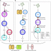
Figure 1.
Figure 1.
Scheme for immune pathways leading to acute and chronic lung disease after viral infection or allergen challenge. A, For acute allergic disease, allergen exposure leads to production of allergen-specific IgE that is available to participate in allergen-driven FcεRI signaling, leading to activation of mast cells (and perhaps other cell types) with consequent recruitment of Th2 effector cells to the airway. Th2 cell production of IL-4 and/or IL-13 leads to AA Mac differentiation, whereas IL-5 generation leads to eosinophil (Eos) accumulation and further IL-13 production in the airway. The role of macrophages and any mechanism for positive feedback between Th2 cells, macrophages, eosinophils, and cDCs in the allergic response remains uncertain. B, For acute postviral disease, viruses cause AEC and pDC production of type 1 IFN. Subsequent IFNAR signaling leads to upregulation of FcεRI expression on resident lung cDCs. In turn, FcεRI activation by viral antigen and antiviral IgE leads to production of CCL28 and recruitment of CCR10-expressing IL-13-producing Th2 cells to the lung. IL-13 drives differentiation of AEC precursors toward mucous cells (mucous cell metaplasia) and airway smooth muscle cells to become more reactive to contractile agonists (AHR). C, For chronic postviral disease, viral remnants activate APCs and thereby facilitate CD1d-dependent antigen presentation and consequent activation of invariant CD4 natural killer T (NKT) cells. NKT cells then interact directly with lung macrophages via IL-13 production and binding to the IL-13R as well as contact between invariant Vα14 TCR and glycolipid-loaded CD1d. This interaction leads to increased expression of IL-13R and production of IL-13 that drives a positive feedback loop to amplify IL-13 production and alternative activation of macrophages, including Chi3l3/4, Mmp12, Alox12e, Arg1, and Relmα gene expression in mice and CHIT1, MMP12, and ALOX15 in humans. AA Mac = alternatively activated macrophage; AEC = airway epithelial cell; AHR = airway hyperreactivity; APC = antigen-presenting cell; cDC = conventional dendritic cell; IFN = interferon; IFNAR = interferon A receptor; IL-13R = IL-13 receptor; iNKT = invariant natural killer T; pDC = plasmacytoid dendritic cell; Th = T helper; TCR = T-cell receptor. (Modified with permission from Benoit and Holtzman.)

Similar articles

Cited by

References

    1. Mosser DM, Edwards JP. Exploring the full spectrum of macrophage activation. Nat Rev Immunol. 2008;8(12):958–969. - PMC - PubMed
    1. Gordon S, Martinez FO. Alternative activation of macrophages: mechanism and functions. Immunity. 2010;32(5):593–604. - PubMed
    1. Martinez FO, Helming L, Gordon S. Alternative activation of macrophages: an immunologic functional perspective. Annu Rev Immunol. 2009;27:451–483. - PubMed
    1. Holtzman MJ, Byers DE, Benoit LA, et al. Immune pathways for translating viral infection into chronic airway disease. Adv Immunol. 2009;102:245–276. - PMC - PubMed
    1. Walter MJ, Kajiwara N, Karanja P, Castro M, Holtzman MJ. Interleukin 12 p40 production by barrier epithelial cells during airway inflammation. J Exp Med. 2001;193(3):339–351. - PMC - PubMed

Publication types