. 2011 Nov 1;71(21):6764-72.
doi: 10.1158/0008-5472.CAN-11-0691. Epub 2011 Sep 6.

Itraconazole inhibits angiogenesis and tumor growth in non-small cell lung cancer

Affiliations

Itraconazole inhibits angiogenesis and tumor growth in non-small cell lung cancer

Blake T Aftab et al. Cancer Res. .

Abstract

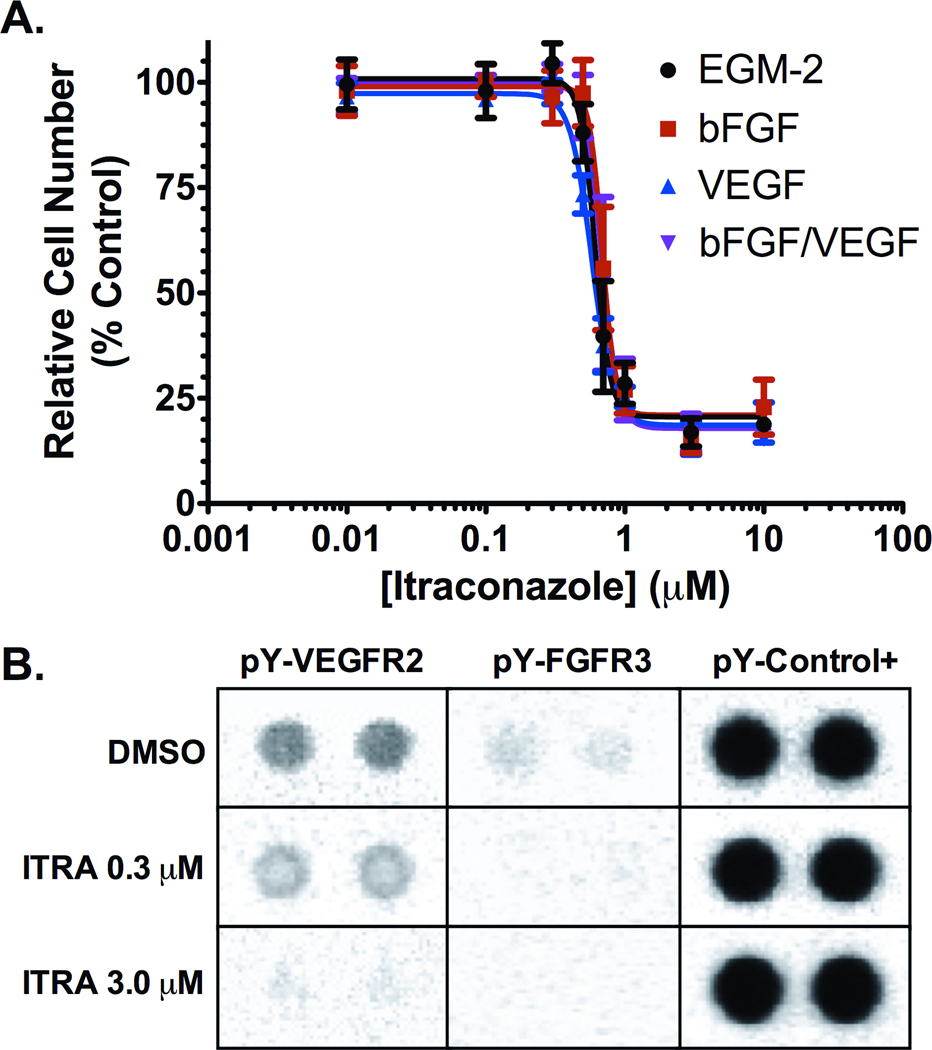
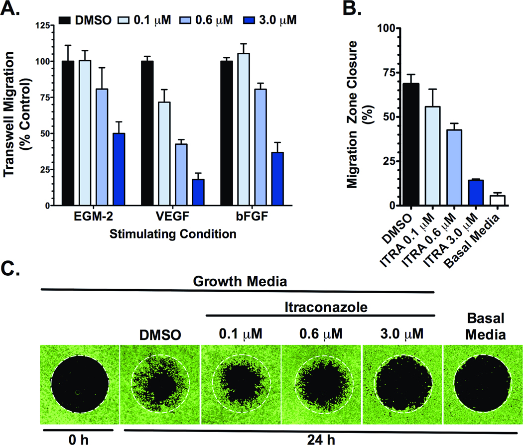
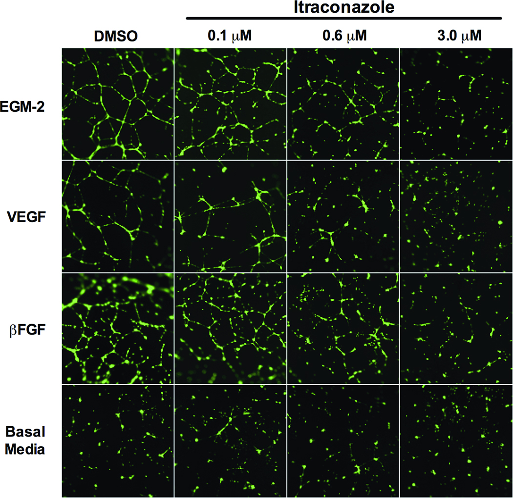
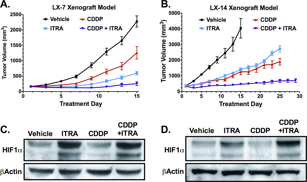
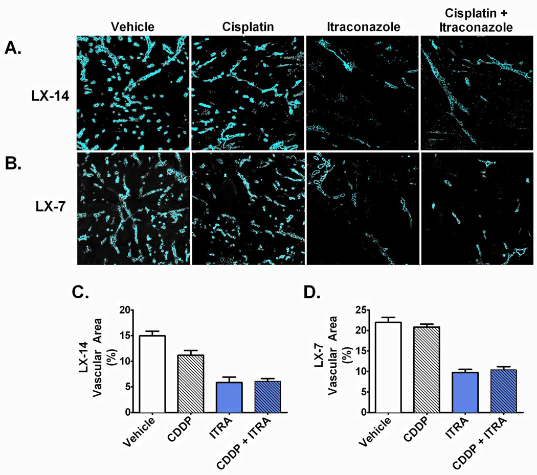
The antiangiogenic agent bevacizumab has been approved for the treatment of non-small cell lung cancer (NSCLC), although the survival benefit associated with this agent is marginal, and toxicities and cost are substantial. A recent screen for selective inhibitors of endothelial cell proliferation identified the oral antifungal drug itraconazole as a novel agent with potential antiangiogenic activity. In this article, we define and characterize the antiangiogenic and anticancer activities of itraconazole in relevant preclinical models of angiogenesis and lung cancer. Itraconazole consistently showed potent, specific, and dose-dependent inhibition of endothelial cell proliferation, migration, and tube formation in response to both VEGF- and basic fibroblast growth factor-mediated angiogenic stimulation. In vivo, using primary xenograft models of human NSCLC, oral itraconazole showed single-agent growth-inhibitory activity associated with induction of tumor hypoxia-inducible factor 1 alpha expression and marked inhibition of tumor vascularity. Itraconazole significantly enhanced the antitumor efficacy of the chemotherapeutic agent cisplatin in the same model systems. Taken together, these data suggest that itraconazole has potent and selective inhibitory activity against multiple key aspects of tumor-associated angiogenesis in vitro and in vivo, and strongly support clinical translation of its use. Based on these observations, we have initiated a randomized phase II study comparing the efficacy of standard cytotoxic therapy with or without daily oral itraconazole in patients with recurrent metastatic NSCLC.


Figures

Figure 1
Figure 1. Inhibition of proliferation and receptor tyrosine kinase phosphorylation in stimulated HUVEC cultures
(A) Itraconazole inhibits HUVEC proliferation in a dose dependent manner in cultures stimulated by supplementation with EGM-2 (black), 12 ng/ml bFGF (red), 10 ng/ml VEGF (blue), and 12 ng/ml bFGF with 10 ng/ml VEGF (purple). Mean relative cell number was evaluated by MTS assay and are reported as mean percent of vehicle treated cell proliferation ± SD. (B) Cell lysates from EGM-2 stimulated HUVEC treated with 0.3 µM or 3.0 µM itraconazole, or with vehicle control for 24 h were hybridized to an RTK array and probed with anti-phospho-tyrosine-HRP antibody followed by chemiluminescent detection. Each RTK is spotted in duplicate with signal normalized to phospho-tyrosine internal controls.
Figure 2
Figure 2. Itraconazole inhibits migration of stimulated HUVEC
(A) HUVEC demonstrate decreased migratory potential across EGM-2, VEGF, and bFGF chemotactic gradients in the presence of increasing concentrations of itraconazole in a transwell assay. Corrected means ± SD of total DNA content from migratory cells are reported as percent of vehicle control for each stimulating condition. (B & C) Itraconazole inhibits EGM-2 stimulated HUVEC migration in a modified wound-healing assay. Following attachment, cells were treated with EGM-2 media containing DMSO vehicle control or 0.1 µM, 0.6 µM, or 3.0 µM itraconazole and permitted to migrate over 24 h into the previously restricted region. Basal media serves as control for unstimulated migration. (B) Migration was quantified as the percent decrease in mean migration zone area ± SD. (C) Representative fluorescence images from the modified wound-healing assay with cells stained by calcein-AM, after a 24 h migration period.
Figure 3
Figure 3. Itraconazole inhibits EGM-2, VEGF, and bFGF stimulated HUVEC tube formation
HUVEC stimulated with EGM-2, 10 ng/ml VEGF, or 12 ng/ml bFGF were allowed to spontaneously form tube networks after treatment with vehicle control, 0.1 µM, 0.6 µM, or 3.0 µM itraconazole. Unstimulated (basal media) HUVEC serve as negative control. Resulting networks were visualized with calcein-AM.
Figure 4
Figure 4. Itraconazole inhibits growth of NSCLC primary xenografts, with itraconazole treated tumors demonstrating increased levels of HIF1α
(A) Mice bearing established LX-7 tumors were treated with vehicle (n=9), oral itraconazole 100 mg/kg bid (ITRA, n=9), cisplatin 4 mg/kg q7d i.p. (CDDP, n=9), or combination itraconazole and cisplatin (ITRA+CDDP, n=8). Mean tumor volume and SEM are reported for each treatment group. (B) Mice bearing established LX-14 tumors were treated with vehicle (n=6), oral itraconazole 100 mg/kg bid (ITRA, n=7), cisplatin 4 mg/kg q7d i.p. (CDDP, n=6), or combination itraconazole and cisplatin (ITRA+CDDP, n=7). Mean tumor volume and SEM are reported for each treatment group. (C) Tumor lysates from LX-7 xenografts were generated from mice treated for 14-days with vehicle (n=4), oral itraconazole (ITRA, n=4), i.p. cisplatin (CDDP, n=4), or combination of itraconazole and cisplatin (ITRA+CDDP, n=4). Pooled lysates were probed for HIF1α. (D) HIF1α immunoblot on pooled tumor lysates from LX-14 xenografts treated for 14-days with vehicle (n=4), oral itraconazole (ITRA, n=4), i.p. cisplatin (CDDP, n=4), or combination of itraconazole and cisplatin (ITRA+CDDP, n=3).
Figure 5
Figure 5. NSCLC tumors demonstrate decreased microvessel density and tumor vascular area following itraconazole treatment
HOE signal from (A) perfused LX-14 tumor and (B) perfused LX-7 sections demonstrate marked decrease in perfusion-competent microvessel density in itraconazole treated tumors. (C & D) Mean ± SEM HOE-positive tumor vascular area in (C) LX-14 tumors treated with vehicle (n=5), itraconazole (ITRA, n=6), cisplatin (CDDP, n=5), or combination itraconazole and cisplatin (ITRA+CDDP, n=7); (D) LX-7 tumors treated with vehicle (n=5), itraconazole (ITRA, n=5), cisplatin (CDDP, n=5), or combination itraconazole and cisplatin (ITRA+CDDP, n=5).

References

    1. Folkman J. Tumor Angiogenesis: Therapeutic Implications. N Engl J Med. 1971;285:1182–1186. - PubMed
    1. Niki T, Iba S, Tokunou M, Yamada T, Matsuno Y, Hirohashi S. Expression of vascular endothelial growth factors A, B, C, and D and their relationships to lymph node status in lung adenocarcinoma. Clin Cancer Res. 2000;6:2431–2439. - PubMed
    1. Han H, Silverman J, Santucci T, Macherey RS, dAmato TA, Tung MY, et al. Vascular Endothelial Growth Factor Expression in Stage I Non-Small Cell Lung Cancer Correlates With Neoangiogenesis and a Poor Prognosis. Ann Surg Oncol. 2001;8:72–79. - PubMed
    1. Bremnes RM, Camps C, Sirera R. Angiogenesis in non-small cell lung cancer: The prognostic impact of neoangiogenesis and the cytokines VEGF and bFGF in tumours and blood. Lung Cancer. 2006;51:143–158. - PubMed
    1. Asahara T, Bauters C, Zheng LP, Takeshita S, Bunting S, Ferrara N, et al. Synergistic effect of vascular endothelial growth factor and basic fibroblast growth factor on angiogenesis in vivo. Circulation. 1995;92:365–371. - PubMed

Publication types

MeSH terms